
Most read articles
- Page Path
- HOME > Browse articles > Most read articles
Most-read are based on citations from 2024 ~ 2026.
Case Report
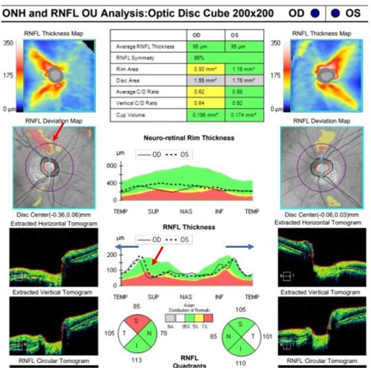
- Delayed toxic anterior segment syndrome after cataract surgery: a case report
- Yeoun Sook Chun
- Insights Cataract Refract Surg 2025;10(1):26-31. Published online February 28, 2025
- DOI: https://doi.org/10.63375/icrs.25.005
-
 Abstract
 PDF
PDF ePub
ePub - Purpose
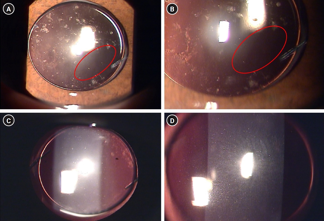
This report describes an unusual case of delayed toxic anterior segment syndrome (TASS) following cataract surgery and its treatment.
Case
summary: A 55-year-old male patient underwent uneventful phacoemulsification with implantation of an intraocular lens (IOL) and eye patching with ophthalmic ointment at the end of the operation. At 1 week postoperatively, a significant increase in the number of anterior chamber inflammatory cells and multiple gray-white deposits on the anterior surface of IOL were noted. All laboratory tests to exclude infectious endophthalmitis were negative. Under the presumptive diagnosis of delayed TASS, an intensive topical steroid was administered. The number of anterior chamber cells decreased; however, the patient complained of blurry vision and multiple whitish precipitates remained on the IOL. Neodymium:yttrium-aluminum-garnet (Nd:YAG) laser treatment was performed to disrupt and remove the precipitates. The deposits were easily and clearly removed using the laser, and there was no recurrence during a 2-year follow-up.
Conclusion
Delayed-onset TASS can manifest as lumpy white inflammatory cell deposits that cannot be controlled with topical steroids. However, Nd:YAG laser treatment can effectively remove inflammatory precipitates.
- 4,043 View
- 34 Download

Editorial
- Personal history of the silicone phakic posterior chamber intraocular lens
- Kenneth J Hoffer
- Insights Cataract Refract Surg 2025;10(1):2-6. Published online February 28, 2025
- DOI: https://doi.org/10.63375/icrs.25.001
- 3,499 View
- 37 Download
Case Report
- Diffuse lamellar keratitis after small incision lenticule extraction: presumably related to meibomian gland dysfunction
- Sang Beom Han
- Insights Cataract Refract Surg 2025;10(2):61-64. Published online June 30, 2025
- DOI: https://doi.org/10.63375/icrs.25.008
-
Abstract
PDFePub
- Purpose
This report presents a case of diffuse lamellar keratitis (DLK) after femtosecond laser-assisted small incision lenticule extraction (SMILE). The case was presumably associated with meibomian gland dysfunction (MGD).
Case
summary: A 25-year-old male patient underwent SMILE surgery. Preoperative examination revealed MGD in both eyes. Despite vigorous cleaning of the eyelid margin and irrigation of the ocular surface, meibomian gland secretion floating on the ocular surface was observed after the lenticule extraction in the right eye. At 2 days postoperatively, stage I DLK was detected. After aggressive topical steroid treatment, the DLK completely resolved without any sequalae.
Conclusion
DLK can occur in association with MGD. Attention should be paid when performing SMILE in eyes with MGD.
- 3,298 View
- 28 Download
Original Article
- Visual and refractive outcomes of keratorefractive lenticule extraction using VISUMAX 800 (SMILE Pro) to correct myopia in Koreans: a 3-month follow-up study
- Sang-Mok Lee, Si-Hoon Park, Tae Keun Yoo, Jae Hyoung Park, Beom Jin Cho, Kee Yong Choi, Jong Woo Kim
- Insights Cataract Refract Surg 2025;10(1):13-18. Published online February 28, 2025
- DOI: https://doi.org/10.63375/icrs.25.004

-
Abstract
PDFePub
- Purpose
The aim of this study was to report the clinical outcomes of SMILE Pro surgery in Korean myopia patients.
Methods
A retrospective analysis was conducted on the medical records of 90 patients (178 eyes) who underwent SMILE Pro surgery at our institution between October 2023 and June 2024 and were followed for 3 months postoperatively.
Results
Preoperative best corrected visual acuity was 0.009±0.020 (logarithm of the minimum angle of resolution). The average spherical equivalent was –5.13±2.16 diopters (range, –1.00 to –10.10 diopters), and the average astigmatism was –1.21±0.91 diopters (range, 0 to –4.0 diopters). Postoperatively, the uncorrected distance visual acuity at 1 day, 1 week, 1 month, and 3 months were 0.061±0.054, 0.013±0.027, 0.009±0.023, 0.005±0.021, respectively. At 3 months postsurgery, the predictive accuracy for spherical equivalent was 100% within ±0.5 diopters and 98.9% within ±0.25 diopters. For astigmatism, the predictive accuracy was 97.2% within ±0.25 diopters and 99.4% within ±0.5 diopters 97.2% and 99.4%. The scores for the efficacy and safety of refractive surgery at 3 months were both 1.01±0.05.
Conclusion
SMILE Pro surgery for myopia correction in Korean patients demonstrated excellent efficacy, safety, and predictive accuracy, with no significant difference compared to conventional SMILE surgery.
- 2,302 View
- 24 Download
Review Articles
- Selection of an optimal intraocular lens according to the stage of epiretinal membrane
- Sang Beom Han
- Insights Cataract Refract Surg 2025;10(1):7-12. Published online February 28, 2025
- DOI: https://doi.org/10.63375/icrs.25.003
-
Abstract
PDFePub
- Epiretinal membrane (ERM), one of the most common retinal diseases, can cause various degrees of visual disturbance, reduced contrast sensitivity, and metamorphopsia. ERM is not infrequently encountered during preoperative evaluations for cataract surgery, and selecting an appropriate intraocular lens (IOL) according to the location and stage of ERM is necessary in order to improve visual outcomes and patients’ satisfaction. This review summarizes the application of various IOLs—such as multifocal, extended depth of focus, and enhanced monofocal IOLs—in eyes with ERM, and discusses the selection of an appropriate IOL.
-
Citations
Citations to this article as recorded by
- Presbyopia-correcting intraocular lens options in myopic eyes undergoing cataract surgery
Sang Beom Han
Insights in Cataract and Refractive Surgery.2026; 11(1): 1. CrossRef
- Presbyopia-correcting intraocular lens options in myopic eyes undergoing cataract surgery
- 1,804 View
- 19 Download
- 1 Crossref
- Current updates in the treatment of keratoconus
- Mee Kum Kim
- Insights Cataract Refract Surg 2025;10(2):33-41. Published online June 30, 2025
- DOI: https://doi.org/10.63375/icrs.25.010
-
Abstract
PDFePub
- Corneal collagen cross-linking is a primary treatment to slow or halt the progression of keratoconus. For visual rehabilitation, important treatments include contact lenses fitting, intracorneal ring segment implantation, and corneal transplantation. Additionally, either phakic intraocular lenses or corneal therapeutic keratectomy combined with corneal collagen cross-linking can assist in visual rehabilitation with limited indications. New methods for visual rehabilitation, such as stromal keratophakia, have been introduced. This review evaluates and compares the efficacy and safety of various treatments for keratoconus based on the latest information.
- 1,694 View
- 27 Download
Original Article
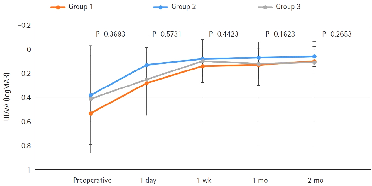
- Efficacy of extended depth of focus, enhanced monofocal, and monofocal intraocular lenses in patients with retinal disease
- Eun Chul Kim
- Insights Cataract Refract Surg 2025;10(2):52-60. Published online June 30, 2025
- DOI: https://doi.org/10.63375/icrs.25.009
-
Abstract
PDFePub
- Purpose
The aim of this study was to compare the visual quality of extended depth of focus (EDOF), enhanced monofocal, and monofocal intraocular lenses (IOLs) in patients with retinal disease.
Methods
In total, 103 eyes from 93 patients (group 1: enhanced monofocal ICB00, n=36; group 2: EDOF ZXR00, n=36; group 3: monofocal ZCB00, n=31) were retrospectively enrolled. Uncorrected and corrected near visual acuity (UNVA, CNVA), intermediate visual acuity (UIVA, CIVA), and distance visual acuity (UDVA, CDVA), manifest refraction spherical equivalent (MRSE), and satisfaction scores were assessed before and after surgery.
Results
The postoperative UDVA, CDVA, and MRSE of the three groups were better than the preoperative data, respectively (P<0.05). The UIVA of group 1 (0.13±0.12 logMAR) and 2 (0.10±0.11) was significantly better than that of groups 3 (0.25±0.15) (P<0.05). The UNVA of group 2 (0.18±0.12) was significantly better than that of groups 1 (0.32±0.20) and 3 (0.45±0.26; P<0.05). The UDVA of patients with macular edema and macular holes was insignificantly lower than that of epiretinal membranes and high myopia. The overall satisfaction of group 1 (1.58±0.81) and 2 (1.46±0.75) was significantly better than that of groups 3 (1.83±0.97; P<0.05).
Conclusion
EDOF and enhanced monofocal IOLs were associated with better intermediate and near vision than monofocal IOLs in patients with retinal disease. However, monofocal IOLs are recommended in patients with macular edema and macular holes, unlike patients with epiretinal membranes and high myopia.
- 1,559 View
- 21 Download
Review Article
- Trabecular microbypass using iStent combined with cataract surgery
- Yeoun Sook Chun
- Insights Cataract Refract Surg 2025;10(2):42-51. Published online June 30, 2025
- DOI: https://doi.org/10.63375/icrs.25.007
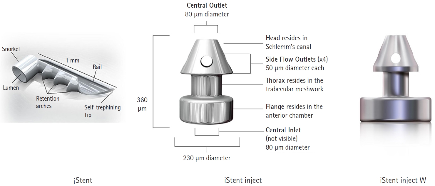
-
Abstract
PDFePub
- Minimally invasive glaucoma surgery has revolutionized conventional glaucoma treatment due to its simple procedures, rapid recovery, and few complications. iStent, a trabecular microbypass that can be implanted via combined cataract surgery, has the advantage of lowering intraocular pressure (IOP) independent of bleb formation. Furthermore, it is straightforward to implement and does not involve a substantial burden. iStent has a synergetic effect with phacoemulsification. Combining iStent with phacoemulsification yielded a greater reduction of IOP and glaucoma eyedrops, as well as higher rates of visual field change, than when either modality was used in isolation. iStent has emerged as a new treatment option for patients with mild to moderate glaucoma. This review aims to improve readers’ understanding of iStent by summarizing the surgical techniques needed to correctly insert it for optimal outcomes and discussing problem-solving in the context of patient care.
- 1,503 View
- 10 Download
Original Article
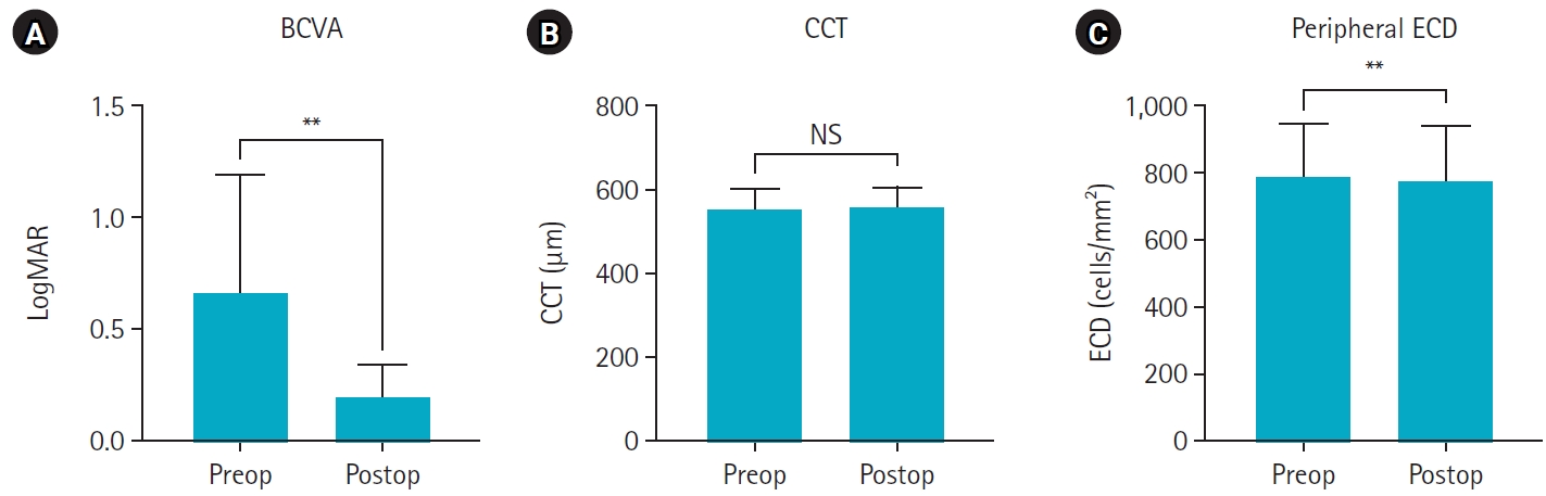
- Clinical manifestations after cataract surgery in patients with moderate Fuchs corneal endothelial dystrophy
- Myung-Sun Song, Dong Hyun Kim
- Insights Cataract Refract Surg 2025;10(1):19-25. Published online February 28, 2025
- DOI: https://doi.org/10.63375/icrs.25.006
-
Abstract
PDFePub
- Purpose
The aim of this study was to analyze the clinical outcomes of cataract surgery in patients with moderate Fuchs endothelial corneal dystrophy (FECD) in whom central endothelial cells could not be observed using specular microscopy.
Methods
This retrospective study included nine eyes in seven patients diagnosed with FECD who underwent phacoemulsification at a single institution between January 2023 and November 2024. A single experienced corneal specialist performed slit-lamp examination and phacoemulsification. Best-corrected visual acuity (BCVA), specular microscopy, and central corneal thickness (CCT) measurements were performed preoperatively and postoperatively, and the outcomes were compared.
Results
The mean age of the patients was 69.8±6.5 years. Three were male patients and four were female patients. The mean preoperative CCT was 559.5±51.8 μm and the mean peripheral endothelial cell density was 599.3±129.4 cells/mm2. BCVA significantly improved in all patients postoperatively, with a mean logMAR BCVA improving from 0.65±0.52 preoperatively to 0.19±0.14 postoperatively (P=0.002). The mean CCT showed no significant change (preoperative, 559.6±51.8 μm; postoperative, 566.8±45.1 μm; P=0.218). In patients with follow-up longer than 6 months, an increase in CCT was observed at 30 days postoperatively, but CCT returned to preoperative levels after 90 days.
Conclusion
In patients with moderate-to-severe FECD in whom central endothelial cells cannot be measured, phacoemulsification may provide favorable visual outcomes if peripheral endothelial cells are observed and corneal edema is absent preoperatively.
- 1,340 View
- 14 Download
Review Article
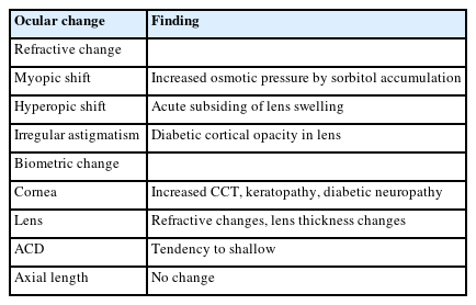
- Phacoemulsification in patients with diabetes: from preoperative evaluation to postoperative management
- Yeoun Sook Chun
- Insights Cataract Refract Surg 2025;10(3):65-75. Published online October 31, 2025
- DOI: https://doi.org/10.63375/icrs.25.012
-
Abstract
PDFePub
- Diabetes mellitus is one of the most common chronic diseases worldwide and is a leading cause of blindness in patients over the age of 50 years. Patients with diabetes have an elevated risk of developing cataracts compared to individuals without diabetes; furthermore, cataracts also tend to progress more rapidly in this population, leading to the need for surgery at a younger age. This review aims to summarize the key considerations in the management of cataract surgery in patients with diabetes, from preoperative evaluation to postoperative care. Patients with diabetes often present with unstable refractive status, dry eye disease, corneal epithelial defects, and recurrent corneal erosions. They also tend to have reduced corneal endothelial cell density and small pupils, both of which increase the risk of intraoperative complications. Postoperatively, these patients are at risk of developing pseudophakic cystoid macular edema, posterior capsular opacification, endophthalmitis, progression of diabetic retinopathy, and neovascular glaucoma. Patients with long-standing or poorly controlled diabetes face a higher likelihood of postoperative complications, highlighting the importance of regular ophthalmic follow-up examinations. Furthermore, adjunctive treatments such as timely intravitreal injections of anti-vascular endothelial growth factor agents may reduce the risk of vision-threatening complications following cataract surgery.
- 1,253 View
- 14 Download
Original Article
- The effect of 2% rebamipide ophthalmic solution on early dry eye after SMILE surgery: a retrospective study
- Jin Hyoung Kim, Mu Yan Kim, Young A Kwon, Sung Wook Park, Sung Won Byun, Seong Woo Lee, Sung Hyup Lim
- Insights Cataract Refract Surg 2025;10(3):91-102. Published online October 31, 2025
- DOI: https://doi.org/10.63375/icrs.25.013
-
Abstract
PDFePub
- Purpose
The aim of this study was to investigate the effect of rebamipide 2% ophthalmic solution on early dry eye following small incision lenticule extraction (SMILE) surgery by analyzing dry eye indicators before and after the procedure.
Methods
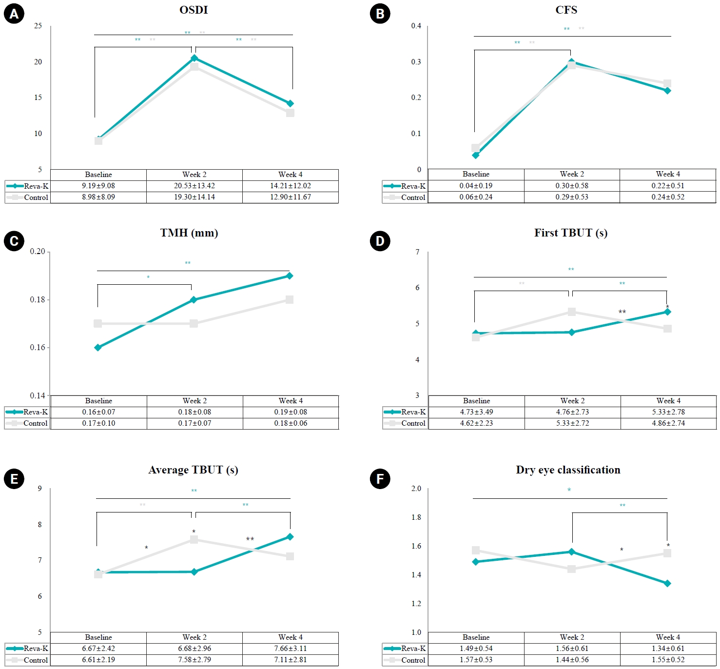
In this retrospective study, an initial sample of 372 SMILE surgery patients were divided into a rebamipide group (artificial tears and 2% rebamipide) and a control group (artificial tears only). Changes in dry eye indicators, including the Ocular Surface Disease Index (OSDI), corneal fluorescein staining (CFS) score, tear meniscus height (TMH), tear break-up time (TBUT), and dry eye classification, were analyzed at 2 and 4 weeks postoperatively in comparison with the preoperative baseline.
Results
In total, 250 patients (250 eyes) were selected: 135 in the rebamipide group and 115 in the control group. Preoperative characteristics such as gender, age, spherical equivalent refraction, ablation depth, and optical zone size showed no significant differences between the two groups. Both groups demonstrated a significant increase in OSDI and CFS scores at 2 weeks postoperatively, followed by a decrease at 4 weeks, with no significant differences between groups. TMH increased significantly in the rebamipide group at 2 and 4 weeks (P=0.043, P=0.004), but showed no significant change in control group or intergroup difference. No significant difference was found in the first TBUT between the two groups at any time point, but the average TBUT significantly and rapidly increased from 2 to 4 weeks postoperatively in the rebamipide group (P=0.001). The dry eye classification was significantly lower in the rebamipide group at 4 weeks postoperatively (P=0.014).
Conclusion
The use of rebamipide 2% ophthalmic solution immediately after SMILE surgery is expected to be helpful in treating early postoperative dry eye, as it increased TMH and TBUT starting from 2 weeks postoperatively.
- 1,106 View
- 10 Download
Editorial
- Insights in Cataract and Refractive Surgery: a new beginning for a leap forward
- Jong Suk Song
- Insights Cataract Refract Surg 2025;10(1):1-1. Published online February 28, 2025
- DOI: https://doi.org/10.63375/icrs.25.002
- 997 View
- 12 Download
Original Articles
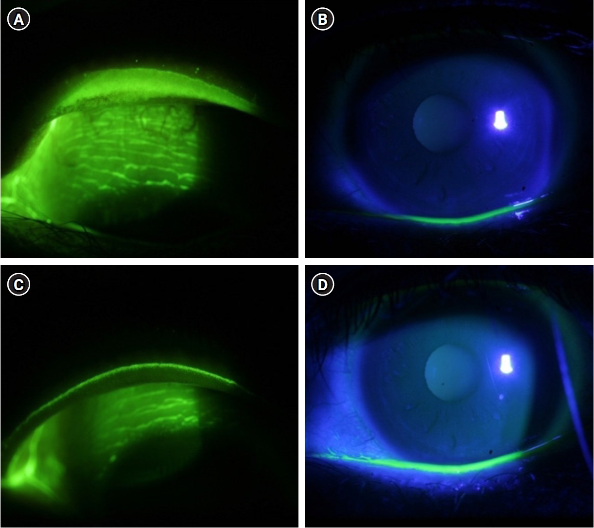
- Therapeutic effects of diquafosol ophthalmic solution and carbomer eye gel in dry eye patients with lid wiper epitheliopathy
- Jong Suk Song, In Ho Woo
- Insights Cataract Refract Surg 2026;11(1):9-14. Published online February 26, 2026
- DOI: https://doi.org/10.63375/icrs.25.019
-
Abstract
PDFePub
- Purpose
This study aimed to evaluate the therapeutic effects of 3% diquafosol tetrasodium ophthalmic solution (Diquas) and a carbomer-based, lipid-containing eye gel (Liposic, Bausch & Lomb) in dry eye patients with lid wiper epitheliopathy (LWE) that was refractory to topical treatment with conventional artificial tears.
Methods
Thirty-three dry eye patients with LWE of the upper eyelid were treated with 3% diquafosol ophthalmic solution administered six times daily and a carbomer-based, lipid-containing eye gel administered four times daily. After the 2-week treatment period, changes in ocular symptoms were assessed using a visual analog scale (VAS), and changes in ocular signs were evaluated using tear film break-up time (TBUT), corneal staining, and LWE grading.
Results
The mean patient age was 37.58±12.35 years (range, 21–67 years); six patients were male and 27 were female. At baseline, the mean VAS symptom score was 7.18±1.47, and the mean TBUT was 2.78±0.78 seconds. After 2 weeks of treatment, the mean VAS score decreased to 4.87±1.97, and the mean TBUT increased to 3.68±1.08 seconds (both P<0.05). The mean corneal staining score was 1.09±1.50, and the mean LWE grade was 5.76±0.61 at baseline. Following treatment, these values decreased to 0.55±0.83 and 2.24±1.95, respectively (both P<0.05). Among the objective parameters, only the LWE grade showed a significant correlation with the VAS score.
Conclusion
The LWE grade showed a significant correlation with ocular symptoms. Combined treatment with diquafosol ophthalmic solution and a carbomer-based, lipid-containing eye gel demonstrated excellent therapeutic effects in dry eye patients with LWE who were refractory to treatment with conventional artificial tears.
- 920 View
- 11 Download
- Results of multifocal intraocular lens implantation in patients who underwent corneal refractive surgery
- Eun Chul Kim
- Insights Cataract Refract Surg 2025;10(3):76-82. Published online October 31, 2025
- DOI: https://doi.org/10.63375/icrs.25.011
-
Abstract
PDFePub
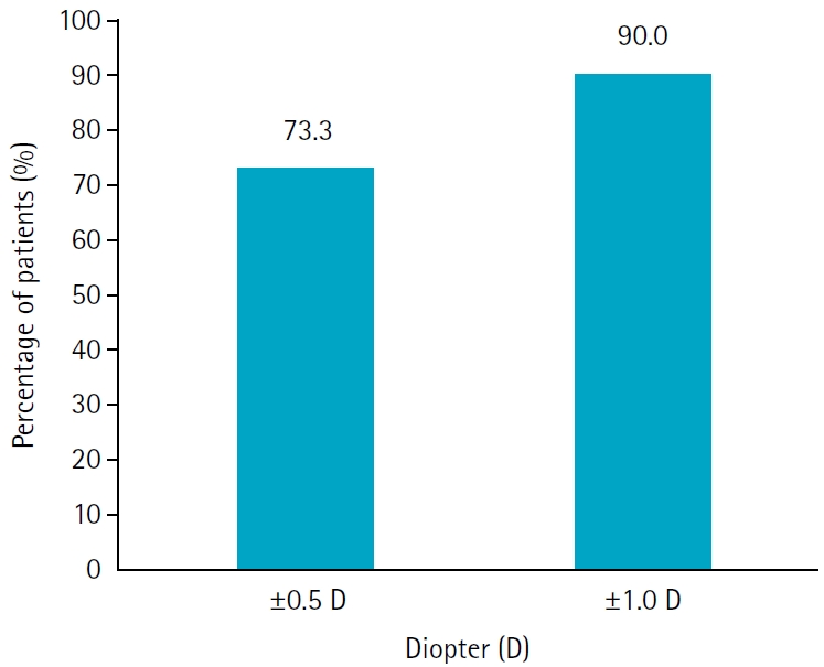
- Purpose
The aim of this study was to evaluate the clinical results of multifocal intraocular lenses in patients who underwent corneal refractive surgery.
Methods
Thirty eyes (16 patients; Synergy: ZFR00V) were retrospectively enrolled. Uncorrected and corrected near visual acuity (UNVA, CNVA), intermediate visual acuity (UIVA, CIVA), and distant visual acuity (UDVA, CDVA), manifest refraction spherical equivalent (MRSE), and satisfaction score were assessed before and after surgery.
Results
The postoperative UDVA, UIVA, UNVA, and MRSE of the three groups exhibited improvements compared to the preoperative data (P<0.05). The error between the postoperative refraction and the intraocular lens calculation was smaller with the Barrett True K formula than with the Haigis-L formula (P<0.05). The defocus curve at 0 diopter (D) increased, from –1 to –1.5 D, and from –2.5 to –4.0 D, indicating improved vision at distant, intermediate, and near distances. Distance satisfaction (1.47±0.63), near satisfaction (1.25±0.71), and overall satisfaction (1.36±0.42) were good, but light scattering and halo satisfaction (1.97±0.85) yielded a poor result.
Conclusion
In patients with cataracts who underwent corneal refractive surgery, multifocal intraocular lens implantation resulted in excellent uncorrected visual acuity at distant, intermediate, and near distances. However, careful consideration should be given to patient selection due to the incidence of side effects such as glare and halos.
- 909 View
- 14 Download
- Clinical results of combined Descemet membrane keratoplasty and cataract operation (triple Descemet membrane keratoplasty) from imported donor corneas: a retrospective study
- Hyung Keun Lee, Sung Soo Kang, Jin Suk Chun, So Young Kim, Dong Ihll Lee
- Insights Cataract Refract Surg 2025;10(3):83-90. Published online October 31, 2025
- DOI: https://doi.org/10.63375/icrs.25.014
-
Abstract
PDFePub
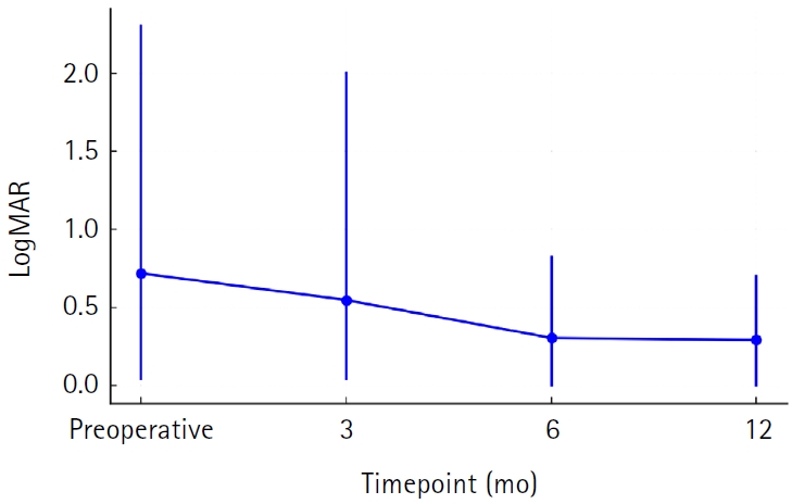
- Purpose
This study reports the clinical outcomes, after triple Descemet membrane endothelial keratoplasty (DMEK) performed using imported corneas.
Methods
A retrospective study was conducted on 30 eyes of 26 patients who underwent Descemet's membrane keratoplasty concurrently with cataract surgery, referred to as triple DMEK, from January 2023 to June 2025. After routine preoperative examinations for keratoplasty as well as cataract surgery, uneventful DMEK surgery was performed concurrently with cataract surgery. All patients visited the clinic at 1, 3, 6, and 12 months after surgery to observe changes, including uncorrected and best spectacle corrected visual acuity, refractive error, corneal thickness, and endothelial cell density.
Results
Preoperatively, 12 of the 30 eyes had Fuchs corneal endothelial dystrophy and 12 had endothelial failure following phakic intraocular lens implantation. The average observation period for the subjects was 437±263 days. After triple DMEK surgery, the patients' uncorrected visual acuity gradually improved from 0.73±0.6 (logMAR) before surgery to 0.65±0.54 at 3 months and 0.29±0.26 at 1 year (P<0.001). The change in corneal thickness was 565.7±70.0 μm before surgery, 535.2±44.2 μm at 3 months after surgery, 549.7±73.5 μm at 6 months, and 535.82±49.0 μm at 12 months, but no statistical significance was found compared to before surgery at any time point (P>0.05). The endothelial cell density was 798±363 cells/mm2 before surgery, 1,479±475 cells/mm2 at 3 months after surgery, 1,456±456 cells/mm2 at 6 months, 1,332±346 cells/mm2 at 12 months, and 1,399±519 cells/mm2 at the last visit (P<0.001).
Conclusion
Triple DMEK surgery, which is performed for various corneal diseases, is relatively safe. No significant endothelial damage, refractive changes, or visual acuity abnormalities were observed up to one year after surgery. Future prospective studies including a larger number of participants are warranted to evaluate the safety and clinical outcomes of triple DMEK using imported corneas.
- 849 View
- 7 Download
Review Article
- Glaucoma evaluation and management in refractive surgery candidates: a review
- Yeoun Sook Chun
- Insights Cataract Refract Surg 2026;11(2):27-39. Published online June 18, 2026
- DOI: https://doi.org/10.63375/icrs.26.002
-
Abstract
PDFePub
- The global prevalence of refractive surgery for myopia has increased substantially; however, definitive guidance regarding its effects on glaucoma assessment and progression remains limited. Although advances in technology and diagnostic instruments have improved the detection and monitoring of glaucoma in patients undergoing refractive surgery, concerns persist regarding postoperative intraocular pressure (IOP) elevation and the inaccuracy of IOP measurements associated with reduced central corneal thickness. Myopia is a well-established risk factor for primary open-angle glaucoma, and this risk may be further increased by various intraoperative and postoperative factors related to refractive surgery. Therefore, thorough preoperative glaucoma screening, along with systematic postoperative follow-up and evaluation, is essential. This review delineates key considerations before refractive surgery and summarizes important clinical issues in eyes that have undergone refractive procedures. Furthermore, it outlines the pathogenesis, mechanisms, and management strategies for postoperative IOP elevation.
- 790 View
- 8 Download
Original Article
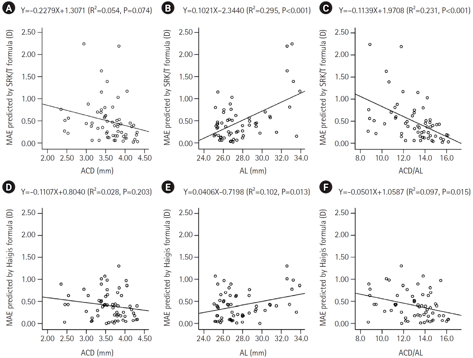
- Impact of anterior chamber depth to axial length ratio on conventional intraocular lens power calculation formulas performance in axial myopia
- Youngsub Eom, Jinhwan Park, Youngbin Song, Dong Hyun Kim, Jong Suk Song
- Insights Cataract Refract Surg 2026;11(1):15-22. Published online February 26, 2026
- DOI: https://doi.org/10.63375/icrs.25.017
-
Abstract
PDFePub
- Purpose
To evaluate the effects of the ratio of anterior chamber depth to axial length (ACD/AL), as well as axial length (AL) itself, on the accuracy of conventional intraocular lens (IOL) power calculation formulas in eyes with axial myopia.
Methods
This retrospective cross-sectional study included 60 eyes from 44 patients with an AL greater than 25.0 mm who underwent uncomplicated phacoemulsification with IOL implantation. Eyes were categorized into high and low AL groups using an AL threshold of 27.0 mm, and into high and low ACD/AL groups based on the median ACD/AL value of 13.4. The median absolute errors (MedAEs) predicted by the Sanders-Retzlaff-Kraff theoretical (SRK/T) and Haigis formulas were compared according to AL and ACD/AL groupings.
Results
In the low ACD/AL group and in the high AL group, the MedAEs predicted by the Haigis formula were lower than those predicted by the SRK/T formula (P=0.002 and P=0.012, respectively). The MedAEs predicted by both the SRK/T and Haigis formulas were significantly lower in the high ACD/AL group than in the low ACD/AL group (P<0.001 and P=0.010, respectively). In contrast, no significant difference was observed between the low and high AL groups in the MedAEs predicted by the Haigis formula. When the ACD/AL ratio was less than 13.4, postoperative refractive outcomes were more hyperopic with both formulas.
Conclusion
In eyes with a long AL and a relatively shallow ACD, the Haigis formula demonstrated superior accuracy among conventional IOL power calculation formulas. Under these anatomical conditions, targeting slightly more myopic postoperative refractions may therefore be advisable.
- 588 View
- 7 Download
Review Article
- Presbyopia-correcting intraocular lens options in myopic eyes undergoing cataract surgery
- Sang Beom Han
- Insights Cataract Refract Surg 2026;11(1):1-8. Published online February 26, 2026
- DOI: https://doi.org/10.63375/icrs.25.016
-
Abstract
PDFePub
- With ongoing advancements in surgical techniques and intraocular lens (IOLs) technologies, cataract surgery is increasingly recognized as a form of refractive procedure aimed at enhancing overall visual performance rather than being viewed solely as lens extraction. In parallel with this shift, a growing number of aging individuals with myopia are actively seeking spectacle independence following cataract surgery. The selection of IOLs for presbyopia correction in patients with myopia presents distinct clinical and refractive challenges, necessitating careful preoperative evaluation and individualized surgical planning. In this review, the author summarizes current evidence regarding the use of various IOLs, including monofocal, enhanced monofocal, extended depth-of-focus, and multifocal IOLs, for presbyopia correction in myopic patients and discusses key considerations involved in selecting the most appropriate IOL for this specific population.
- 556 View
- 8 Download
Case Report
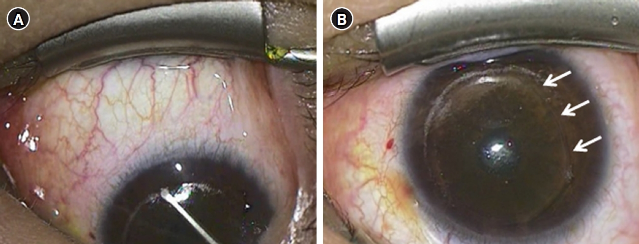
- Large incisional tear caused by abrupt Bell’s phenomenon during pocket irrigation in femtosecond laser-assisted small incision lenticule extraction
- Sang Beom Han
- Insights Cataract Refract Surg 2026;11(1):23-25. Published online February 26, 2026
- DOI: https://doi.org/10.63375/icrs.25.015
-
Abstract
PDFePub
- Purpose
This study reports a case of a large incisional tear caused by abrupt Bell’s phenomenon during pocket irrigation in femtosecond laser-assisted small incision lenticule extraction (SMILE).
Case
summary: A 28-year-old male patient underwent SMILE surgery. During pocket irrigation of the right eye, Bell’s phenomenon suddenly occurred, resulting in a large inferior arcuate extension of the incision that reached the inferior cap margin. After confirming wound integrity and the absence of additional tissue damage, a bandage contact lens was applied. On postoperative day 7, the incision was self-sealed with intact wound integrity. Three months postoperatively, uncorrected distance visual acuity was 20/20 in both eyes. The right cornea remained stable, although a faint residual scar was observed at the site of the incisional tear.
Conclusion
Abrupt eye movements during pocket irrigation in SMILE can cause large incisional tears. Surgeons should exercise heightened vigilance during irrigation, particularly in anxious or uncooperative patients, to prevent such complications.
- 531 View
- 4 Download
Original Articles
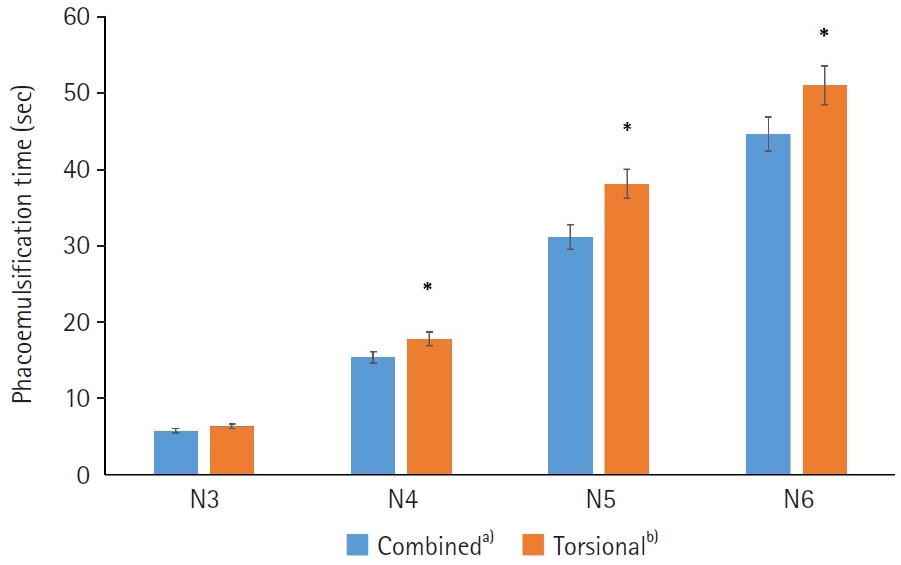
- Comparison of combined torsional and conventional ultrasound mode versus torsional ultrasound mode phacoemulsification in different machines
- Jiyoung Emily Lee, Eun Chul Kim
- Insights Cataract Refract Surg 2026;11(2):40-46. Published online May 21, 2026
- DOI: https://doi.org/10.63375/icrs.25.018
-
Abstract
PDFePub
- Purpose
This study aimed to compare the clinical outcomes of phacoemulsification performed using a combined torsional and conventional ultrasound mode versus torsional ultrasound mode alone in cataract surgery.
Methods
Sixty-five patients who underwent phacoemulsification using combined torsional and conventional ultrasound mode with the Cube alpha system (Nidek Co.) were compared with 300 patients who underwent phacoemulsification using torsional ultrasound mode alone with the Centurion system (Alcon Research Ltd.). The two groups were classified as N3, N4, N5, or N6 according to the Lens Opacities Classification System III. In each subgroup, phacoemulsification time (seconds), cumulative dissipated energy (CDE), balanced salt solution (BSS) use, and postoperative best-corrected visual acuity were analyzed and compared between the combined-mode group and the torsional-only group.
Results
In the N4–N6 subgroups, the torsional-only group had significantly longer phacoemulsification times and higher CDE values than the combined-mode group (P<0.05 for all comparisons). In the N3–N6 subgroups, the total volume of BSS used in the torsional-only group (N3, 30.97±11.05; N4, 48.56±20.06; N5, 68.27±23.06; N6, 125.64±45.67) was significantly greater than that used in the combined-mode group (N3, 23.01±10.27; N4, 39.03±24.16; N5, 42.86±18.24; N6, 54.75±0.75) (P<0.05). There were no statistically significant differences between the groups in postoperative manifest refraction spherical equivalent, corneal endothelial cell loss, or best-corrected visual acuity.
Conclusion
Compared with torsional ultrasound mode alone, the combined torsional and conventional ultrasound mode was more efficient in terms of ultrasound time, CDE, and BSS use. This advantage was more evident with increasing nuclear sclerosis grade.
- 430 View
- 4 Download
- Clinical outcomes of combined phacoemulsification and epiretinal membrane peeling using three advanced intraocular lens platforms
- Chan Hong Min, Jaehyuck Jo, Ho Seok Chung, Dong Yoon Kim, Jin Hyoung Park
- Insights Cataract Refract Surg 2026;11(2):47-55. Published online May 21, 2026
- DOI: https://doi.org/10.63375/icrs.26.001
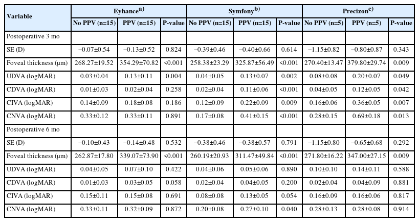
-
Abstract
PDFePub
- Purpose
This retrospective study evaluated the clinical outcomes of combined phacoemulsification, implantation of 1 of 3 types of advanced intraocular lenses (IOLs) with multifocality, and pars plana vitrectomy with epiretinal membrane (ERM) peeling in patients with cataract and ERM. Outcomes were compared with those in age-matched controls who underwent phacoemulsification with the same IOL alone.
Methods
A total of 70 eyes were included: 35 eyes underwent combined surgery, and 35 served as controls. In each group, 15 eyes received an advanced monofocal IOL (Tecnis Eyhance ICB00), 15 received a hybrid diffractive extended-depth-of-focus (EDoF) IOL (Tecnis Symfony ZXR00), and five received a hybrid refractive EDoF IOL (Precizon Presbyopic NVA).
Results
All groups showed significant visual improvement by 6 months postoperatively. Eyes that received hybrid diffractive EDoF IOLs and underwent combined surgery showed significantly worse mean corrected distance visual acuity, uncorrected distance visual acuity, corrected intermediate visual acuity, and corrected near visual acuity at 3 months than control eyes, but these outcomes improved to levels comparable to those of control eyes by 6 months. Eyes that received hybrid refractive EDoF IOLs showed similar early delays; however, interpretation was limited by the small sample size.
Conclusion
Overall, combined surgery was safe and effective, although early visual recovery may be delayed in eyes receiving EDoF IOLs. These findings suggest that advanced monofocal and EDoF IOLs may be considered in selected patients with ERM; however, the results should be interpreted cautiously because of the retrospective design and limited sample size.
- 330 View
- 6 Download
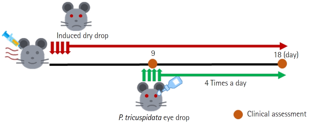
- Effects of extract of Parthenocissus tricuspidata living on pine in a nonclinical model of dry eye disease
- Hyeyoon Goo, Chung-Hun Oh, Kyong Jin Cho
- Insights Cataract Refract Surg 2026;11(2):56-68. Published online June 30, 2026
- DOI: https://doi.org/10.63375/icrs.26.005
-
Abstract
PDF
 Supplementary MaterialePub
 - Purpose
Dry eye disease (DED) is a multifactorial ocular surface disorder characterized by tear film instability, hyperosmolarity, and inflammation. Oxidative stress plays an important role in DED pathogenesis by exacerbating ocular surface damage. Parthenocissus tricuspidata growing on pine (PT) has been reported to have antioxidant and anti-inflammatory properties.
Methods
Oxidative stress was induced in human conjunctival epithelial cells (Wong-Kilbourne derivative of Chang conjunctival [WKD] cells) using H2O2, and the antioxidant and protective effects of PT were evaluated. The anti-inflammatory and therapeutic effects of PT were also investigated in a mouse model of DED.
Results
In WKD cells, PT treatment reduced H2O2-induced apoptosis, reactive oxygen species production, and phosphorylation of mitogen-activated protein kinase signaling proteins. Antioxidant enzyme activity, including superoxide dismutase and catalase, increased, whereas malondialdehyde and interleukin-6 levels decreased, indicating reduced oxidative stress and inflammation. In vivo, PT eye drops significantly improved clinical signs of DED, including tear volume and tear film break-up time. Histological analysis and cytokine assays showed reduced expression of pro-inflammatory markers in corneal and conjunctival tissues.
Conclusion
PT extract exerts therapeutic antioxidant and anti-inflammatory effects, highlighting its potential as a treatment for DED.
- 236 View
- 11 Download
 First
First Prev
Prev

