
 KSCRS
KSCRS
Ahead-of print articles
- Page Path
- HOME > Browse articles > Ahead-of print articles
Articles in E-pub version are posted online ahead of regular printed publication.
Original Article
- Comparison of toric intraocular lenses and limbal relaxing incision in extended-depth-of-focus intraocular lens implantation
- Jiyoung Emily Lee, Eun Chul Kim
- Received March 30, 2026 Accepted August 14, 2026 Published online September 23, 2026
- DOI: https://doi.org/10.63375/icrs.26.003 [Epub ahead of print]
-
 Abstract
 PDF
PDF ePub
ePub - Purpose
This study aimed to compare visual and refractive outcomes after toric intraocular lens (IOL) implantation and limbal relaxing incision (LRI) in eyes receiving extended-depth-of-focus (EDOF) IOLs.
Methods
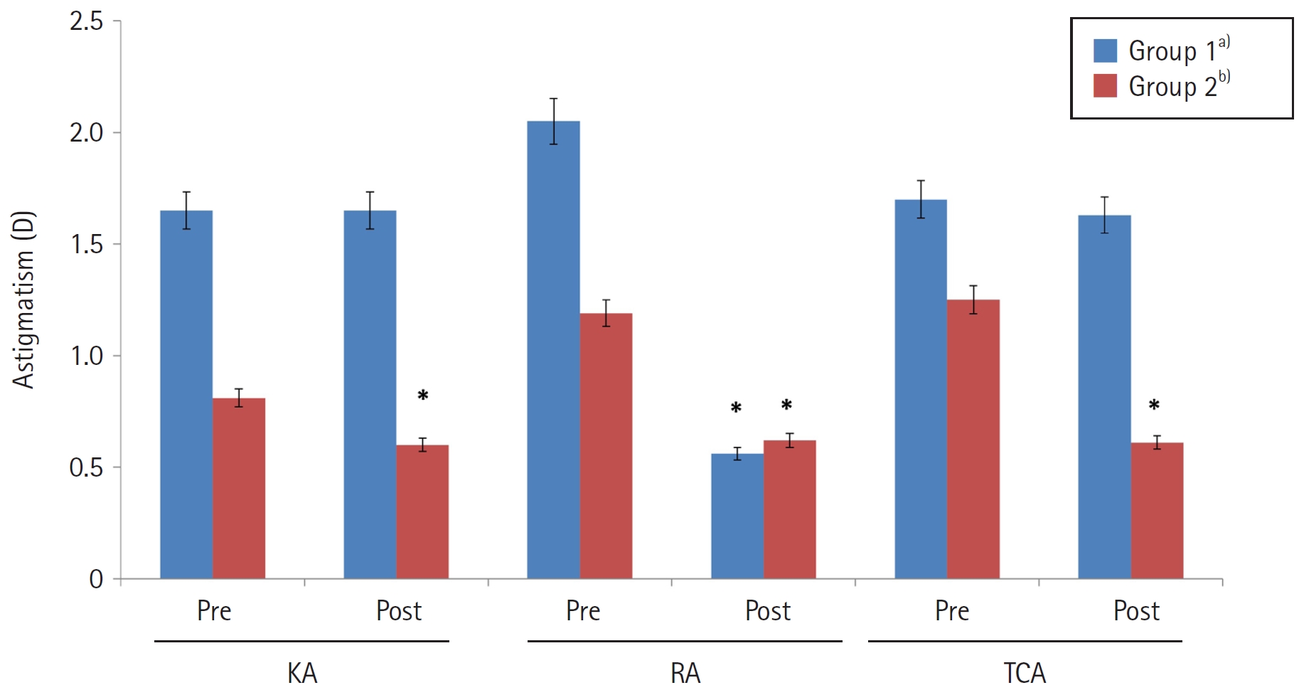
Clinical outcomes were compared between 60 patients who underwent EDOF toric IOL implantation and 60 patients who underwent EDOF IOL implantation combined with LRI. A toric IOL was implanted when corneal astigmatism exceeded 1.0 diopters (D) (group 1), whereas LRI was performed when corneal astigmatism was 1.0 D or less (group 2). Corneal astigmatism, refractive astigmatism, refraction, uncorrected visual acuity, and corrected visual acuity (logMAR) were analyzed in each group before surgery and 3 months postoperatively.
Results
Corneal and total corneal astigmatism did not differ significantly between preoperative and postoperative measurements in group 1 (P>0.05). In group 2, postoperative corneal and total corneal astigmatism (0.60±0.13 and 0.61±0.28 D, respectively) were significantly lower than the corresponding preoperative values (0.81±0.36 and 1.25±0.32 D, respectively; P<0.05). Refractive astigmatism decreased significantly in both group 1 and group 2, from 2.05±0.64 and 1.18±0.41 D preoperatively to 0.56±0.32 and 0.62±0.26 D at 3 months postoperatively, respectively; however, the magnitude of reduction was significantly greater in group 1 than in group 2 (P<0.05). Postoperative refractive astigmatism did not differ significantly between the two groups (P>0.05). In both groups, uncorrected and corrected visual acuity improved significantly after surgery (P<0.05), with no significant between-group difference (P>0.05).
Conclusion
Combining an EDOF IOL with LRI may be effective for mild astigmatism, whereas selecting a toric EDOF IOL may better support visual improvement in patients with moderate to severe astigmatism.
- 109 View
- 0 Download

Case Report
- Therapeutic penetrating keratoplasty in rapidly progressive Acanthamoeba keratitis: a case report
- Yunseo Choi, Jaewon Jung, Tae-im Kim
- Received May 24, 2026 Accepted June 10, 2026 Published online July 31, 2026
- DOI: https://doi.org/10.63375/icrs.26.006 [Epub ahead of print]
-
Abstract
PDFePub
- Purpose
To report the clinical course and postoperative management strategy for emergency therapeutic penetrating keratoplasty (PKP) in a patient with rapidly progressive Acanthamoeba keratitis.
Case
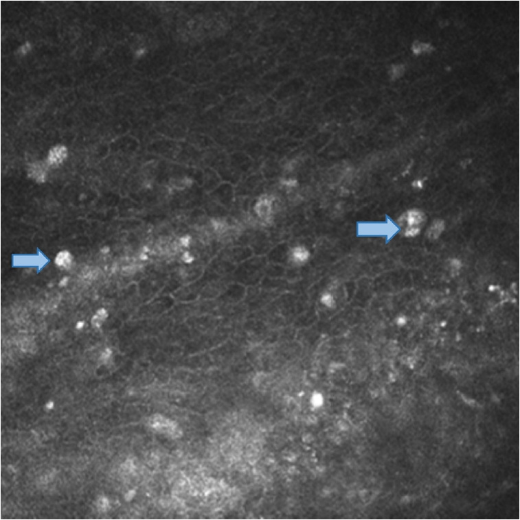
summary: A 23-year-old female patient with a history of long-term orthokeratology lens wear presented with Acanthamoeba keratitis in the right eye. The diagnosis had been established at a referring hospital based on characteristic clinical findings, and topical antiamoebic therapy was initiated, resulting in initial clinical improvement. After polyhexamethylene biguanide was discontinued, an epithelial defect recurred. Amniotic membrane transplantation with adjunctive topical corticosteroids was then performed, after which corneal infiltration and inflammation worsened. At presentation to our clinic, best-corrected visual acuity (BCVA) was light perception, and intraocular pressure was elevated to 34.1 mmHg. Slit-lamp biomicroscopic examination revealed extensive corneal infiltration accompanied by hypopyon. Emergency therapeutic PKP was performed the following day because of uncontrolled infection with severe stromal thinning. Postoperatively, intensive antiamoebic therapy was administered, and topical corticosteroids were initially withheld. Systemic immunosuppression was introduced 1 week postoperatively to manage early graft rejection. Management required careful balancing of immunosuppression for graft survival against the risk of recurrent Acanthamoeba infection. Extensive inferior corneal neovascularization was observed and required additional interventions, including subconjunctival bevacizumab injection. At 5 to 6 months postoperatively, the graft remained clear, with no evidence of recurrent infection, and BCVA had improved to 0.7.
Conclusion
Timely emergency therapeutic PKP with careful postoperative management may serve as a vision-preserving strategy in patients with rapidly progressive Acanthamoeba keratitis.
- 517 View
- 4 Download
 First
First Prev
Prev


