
Search
- Page Path
- HOME > Search
Original Articles
- Clinical outcomes of combined phacoemulsification and epiretinal membrane peeling using three advanced intraocular lens platforms
- Chan Hong Min, Jaehyuck Jo, Ho Seok Chung, Dong Yoon Kim, Jin Hyoung Park
- Insights Cataract Refract Surg 2026;11(2):47-55. Published online May 21, 2026
- DOI: https://doi.org/10.63375/icrs.26.001
-
 Abstract
 PDF
PDF ePub
ePub - Purpose
This retrospective study evaluated the clinical outcomes of combined phacoemulsification, implantation of 1 of 3 types of advanced intraocular lenses (IOLs) with multifocality, and pars plana vitrectomy with epiretinal membrane (ERM) peeling in patients with cataract and ERM. Outcomes were compared with those in age-matched controls who underwent phacoemulsification with the same IOL alone.
Methods
A total of 70 eyes were included: 35 eyes underwent combined surgery, and 35 served as controls. In each group, 15 eyes received an advanced monofocal IOL (Tecnis Eyhance ICB00), 15 received a hybrid diffractive extended-depth-of-focus (EDoF) IOL (Tecnis Symfony ZXR00), and five received a hybrid refractive EDoF IOL (Precizon Presbyopic NVA).
Results
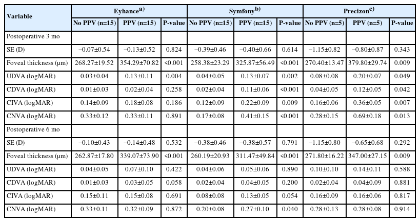
All groups showed significant visual improvement by 6 months postoperatively. Eyes that received hybrid diffractive EDoF IOLs and underwent combined surgery showed significantly worse mean corrected distance visual acuity, uncorrected distance visual acuity, corrected intermediate visual acuity, and corrected near visual acuity at 3 months than control eyes, but these outcomes improved to levels comparable to those of control eyes by 6 months. Eyes that received hybrid refractive EDoF IOLs showed similar early delays; however, interpretation was limited by the small sample size.
Conclusion
Overall, combined surgery was safe and effective, although early visual recovery may be delayed in eyes receiving EDoF IOLs. These findings suggest that advanced monofocal and EDoF IOLs may be considered in selected patients with ERM; however, the results should be interpreted cautiously because of the retrospective design and limited sample size.
- 331 View
- 6 Download

- Clinical results of combined Descemet membrane keratoplasty and cataract operation (triple Descemet membrane keratoplasty) from imported donor corneas: a retrospective study
- Hyung Keun Lee, Sung Soo Kang, Jin Suk Chun, So Young Kim, Dong Ihll Lee
- Insights Cataract Refract Surg 2025;10(3):83-90. Published online October 31, 2025
- DOI: https://doi.org/10.63375/icrs.25.014
-
Abstract
PDFePub
- Purpose
This study reports the clinical outcomes, after triple Descemet membrane endothelial keratoplasty (DMEK) performed using imported corneas.
Methods
A retrospective study was conducted on 30 eyes of 26 patients who underwent Descemet's membrane keratoplasty concurrently with cataract surgery, referred to as triple DMEK, from January 2023 to June 2025. After routine preoperative examinations for keratoplasty as well as cataract surgery, uneventful DMEK surgery was performed concurrently with cataract surgery. All patients visited the clinic at 1, 3, 6, and 12 months after surgery to observe changes, including uncorrected and best spectacle corrected visual acuity, refractive error, corneal thickness, and endothelial cell density.
Results
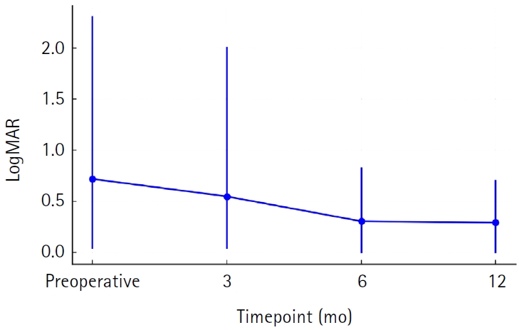
Preoperatively, 12 of the 30 eyes had Fuchs corneal endothelial dystrophy and 12 had endothelial failure following phakic intraocular lens implantation. The average observation period for the subjects was 437±263 days. After triple DMEK surgery, the patients' uncorrected visual acuity gradually improved from 0.73±0.6 (logMAR) before surgery to 0.65±0.54 at 3 months and 0.29±0.26 at 1 year (P<0.001). The change in corneal thickness was 565.7±70.0 μm before surgery, 535.2±44.2 μm at 3 months after surgery, 549.7±73.5 μm at 6 months, and 535.82±49.0 μm at 12 months, but no statistical significance was found compared to before surgery at any time point (P>0.05). The endothelial cell density was 798±363 cells/mm2 before surgery, 1,479±475 cells/mm2 at 3 months after surgery, 1,456±456 cells/mm2 at 6 months, 1,332±346 cells/mm2 at 12 months, and 1,399±519 cells/mm2 at the last visit (P<0.001).
Conclusion
Triple DMEK surgery, which is performed for various corneal diseases, is relatively safe. No significant endothelial damage, refractive changes, or visual acuity abnormalities were observed up to one year after surgery. Future prospective studies including a larger number of participants are warranted to evaluate the safety and clinical outcomes of triple DMEK using imported corneas.
- 850 View
- 7 Download
Review Article
- Phacoemulsification in patients with diabetes: from preoperative evaluation to postoperative management
- Yeoun Sook Chun
- Insights Cataract Refract Surg 2025;10(3):65-75. Published online October 31, 2025
- DOI: https://doi.org/10.63375/icrs.25.012
-
Abstract
PDFePub
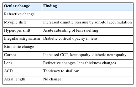
- Diabetes mellitus is one of the most common chronic diseases worldwide and is a leading cause of blindness in patients over the age of 50 years. Patients with diabetes have an elevated risk of developing cataracts compared to individuals without diabetes; furthermore, cataracts also tend to progress more rapidly in this population, leading to the need for surgery at a younger age. This review aims to summarize the key considerations in the management of cataract surgery in patients with diabetes, from preoperative evaluation to postoperative care. Patients with diabetes often present with unstable refractive status, dry eye disease, corneal epithelial defects, and recurrent corneal erosions. They also tend to have reduced corneal endothelial cell density and small pupils, both of which increase the risk of intraoperative complications. Postoperatively, these patients are at risk of developing pseudophakic cystoid macular edema, posterior capsular opacification, endophthalmitis, progression of diabetic retinopathy, and neovascular glaucoma. Patients with long-standing or poorly controlled diabetes face a higher likelihood of postoperative complications, highlighting the importance of regular ophthalmic follow-up examinations. Furthermore, adjunctive treatments such as timely intravitreal injections of anti-vascular endothelial growth factor agents may reduce the risk of vision-threatening complications following cataract surgery.
- 1,254 View
- 14 Download
Case Report
- Delayed toxic anterior segment syndrome after cataract surgery: a case report
- Yeoun Sook Chun
- Insights Cataract Refract Surg 2025;10(1):26-31. Published online February 28, 2025
- DOI: https://doi.org/10.63375/icrs.25.005
-
Abstract
PDFePub
- Purpose
This report describes an unusual case of delayed toxic anterior segment syndrome (TASS) following cataract surgery and its treatment.
Case
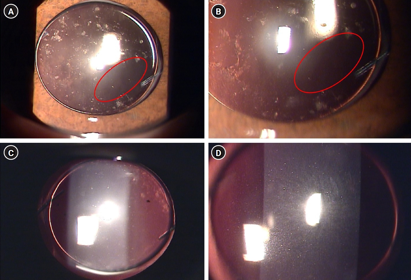
summary: A 55-year-old male patient underwent uneventful phacoemulsification with implantation of an intraocular lens (IOL) and eye patching with ophthalmic ointment at the end of the operation. At 1 week postoperatively, a significant increase in the number of anterior chamber inflammatory cells and multiple gray-white deposits on the anterior surface of IOL were noted. All laboratory tests to exclude infectious endophthalmitis were negative. Under the presumptive diagnosis of delayed TASS, an intensive topical steroid was administered. The number of anterior chamber cells decreased; however, the patient complained of blurry vision and multiple whitish precipitates remained on the IOL. Neodymium:yttrium-aluminum-garnet (Nd:YAG) laser treatment was performed to disrupt and remove the precipitates. The deposits were easily and clearly removed using the laser, and there was no recurrence during a 2-year follow-up.
Conclusion
Delayed-onset TASS can manifest as lumpy white inflammatory cell deposits that cannot be controlled with topical steroids. However, Nd:YAG laser treatment can effectively remove inflammatory precipitates.
- 4,043 View
- 34 Download
 First
First Prev
Prev

