-
Comparison of combined torsional and conventional ultrasound mode versus torsional ultrasound mode phacoemulsification in different machines
-
Jiyoung Emily Lee, Eun Chul Kim
-
Insights Cataract Refract Surg 2026;11(2):40-46. Published online May 21, 2026
-
DOI: https://doi.org/10.63375/icrs.25.018
-
-
 Abstract Abstract
 PDF PDF ePub ePub
- Purpose
This study aimed to compare the clinical outcomes of phacoemulsification performed using a combined torsional and conventional ultrasound mode versus torsional ultrasound mode alone in cataract surgery.
Methods
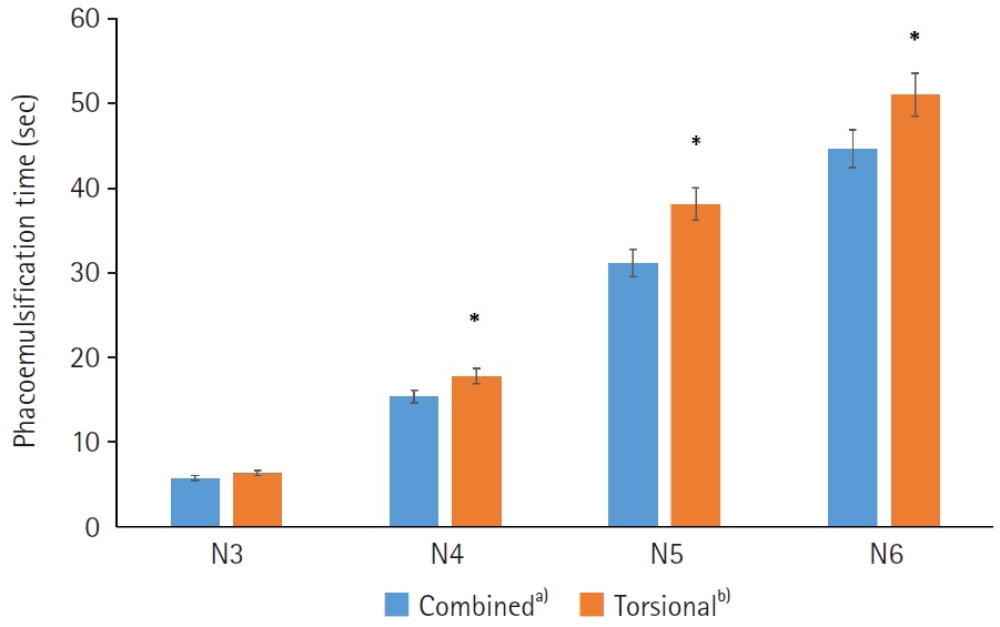
Sixty-five patients who underwent phacoemulsification using combined torsional and conventional ultrasound mode with the Cube alpha system (Nidek Co.) were compared with 300 patients who underwent phacoemulsification using torsional ultrasound mode alone with the Centurion system (Alcon Research Ltd.). The two groups were classified as N3, N4, N5, or N6 according to the Lens Opacities Classification System III. In each subgroup, phacoemulsification time (seconds), cumulative dissipated energy (CDE), balanced salt solution (BSS) use, and postoperative best-corrected visual acuity were analyzed and compared between the combined-mode group and the torsional-only group.
Results
In the N4–N6 subgroups, the torsional-only group had significantly longer phacoemulsification times and higher CDE values than the combined-mode group (P<0.05 for all comparisons). In the N3–N6 subgroups, the total volume of BSS used in the torsional-only group (N3, 30.97±11.05; N4, 48.56±20.06; N5, 68.27±23.06; N6, 125.64±45.67) was significantly greater than that used in the combined-mode group (N3, 23.01±10.27; N4, 39.03±24.16; N5, 42.86±18.24; N6, 54.75±0.75) (P<0.05). There were no statistically significant differences between the groups in postoperative manifest refraction spherical equivalent, corneal endothelial cell loss, or best-corrected visual acuity.
Conclusion
Compared with torsional ultrasound mode alone, the combined torsional and conventional ultrasound mode was more efficient in terms of ultrasound time, CDE, and BSS use. This advantage was more evident with increasing nuclear sclerosis grade.
-
Comparison of toric intraocular lenses and limbal relaxing incision in extended-depth-of-focus intraocular lens implantation
-
Jiyoung Emily Lee, Eun Chul Kim
-
Received March 30, 2026 Accepted August 14, 2026 Published online September 23, 2026
-
DOI: https://doi.org/10.63375/icrs.26.003
[Epub ahead of print]
-
-
Abstract
PDFePub
- Purpose
This study aimed to compare visual and refractive outcomes after toric intraocular lens (IOL) implantation and limbal relaxing incision (LRI) in eyes receiving extended-depth-of-focus (EDOF) IOLs.
Methods
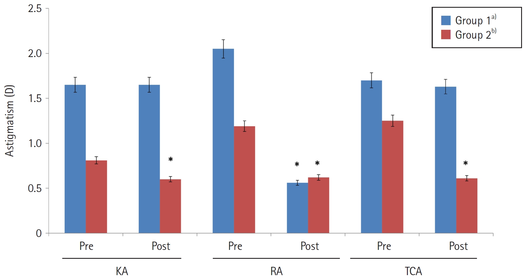
Clinical outcomes were compared between 60 patients who underwent EDOF toric IOL implantation and 60 patients who underwent EDOF IOL implantation combined with LRI. A toric IOL was implanted when corneal astigmatism exceeded 1.0 diopters (D) (group 1), whereas LRI was performed when corneal astigmatism was 1.0 D or less (group 2). Corneal astigmatism, refractive astigmatism, refraction, uncorrected visual acuity, and corrected visual acuity (logMAR) were analyzed in each group before surgery and 3 months postoperatively.
Results
Corneal and total corneal astigmatism did not differ significantly between preoperative and postoperative measurements in group 1 (P>0.05). In group 2, postoperative corneal and total corneal astigmatism (0.60±0.13 and 0.61±0.28 D, respectively) were significantly lower than the corresponding preoperative values (0.81±0.36 and 1.25±0.32 D, respectively; P<0.05). Refractive astigmatism decreased significantly in both group 1 and group 2, from 2.05±0.64 and 1.18±0.41 D preoperatively to 0.56±0.32 and 0.62±0.26 D at 3 months postoperatively, respectively; however, the magnitude of reduction was significantly greater in group 1 than in group 2 (P<0.05). Postoperative refractive astigmatism did not differ significantly between the two groups (P>0.05). In both groups, uncorrected and corrected visual acuity improved significantly after surgery (P<0.05), with no significant between-group difference (P>0.05).
Conclusion
Combining an EDOF IOL with LRI may be effective for mild astigmatism, whereas selecting a toric EDOF IOL may better support visual improvement in patients with moderate to severe astigmatism.
|

 KSCRS
KSCRS