Abstract
-
Purpose
- To describe recurrent keratoconjunctivitis in a patient with X-linked agammaglobulinemia (XLA) and report the findings of microbiome analysis.
-
Case summary
- A 15-year-old male with XLA presented with photophobia, epiphora, redness, and reduced vision in the right eye. Examination revealed corneal infiltration, neovascularization (NV), and thinning in the right eye, with multiple subepithelial opacities in the left. The corneal lesions improved transiently with topical corticosteroids and antibiotics but repeatedly worsened approximately 1 week before each scheduled monthly intravenous immunoglobulin (IVIG) infusion. Approximately 23 months later, corneal NV, central opacity, and thinning also developed in the left eye. Cultures of both conjunctivae yielded Haemophilus influenzae; a right eyelid culture also yielded Staphylococcus epidermidis. Conjunctival microbiome analysis confirmed H. influenzae and detected additional organisms—Mycoplasma amphoriforme, Streptococcus pneumoniae, and Staphylococcus aureus.
-
Conclusion
- In this patient with XLA, topical therapy provided only transient improvement in recurrent keratoconjunctivitis. Exacerbations were temporally associated with the presumed trough period before each scheduled IVIG infusion. Conjunctival microbiome analysis detected additional organisms not identified by bacterial culture.
-
Keywords: Agammaglobulinemia tyrosine kinase; Bruton type agammaglobulinemia; Corneal neovascularization; Keratoconjunctivitis; Microbiome
Introduction
X-linked agammaglobulinemia (XLA) is a very rare inherited immunodeficiency, occurring in approximately 1 per 100,000 to 200,000 live births, and is the most common genetic disorder causing arrest of B-cell maturation [1]. According to the Rare Disease Registry Statistics published by the Korea Disease Control and Prevention Agency [2], four patients were diagnosed with XLA in 2019, three in 2020, two in 2021, two in 2022, and one in 2023 in Korea. XLA is caused by mutations in Bruton's tyrosine kinase (BTK), a key protein involved in signaling downstream of the pre-B-cell receptor and the B-cell receptor [3]. BTK mutations impair precursor B-cell differentiation and result in profound immunodeficiency.
Because of B-lymphocyte deficiency and hypogammaglobulinemia, patients with XLA experience recurrent infections involving multiple organ systems, including upper respiratory tract infections such as otitis media and sinusitis, chronic diarrhea, conjunctivitis, and skin infections [4]. They are also vulnerable to severe and potentially life-threatening infections, including pneumonia, empyema, meningitis, sepsis, cellulitis, and septic arthritis [1,4]. Common bacterial pathogens reported in XLA include Haemophilus influenzae, Streptococcus pneumoniae, Staphylococcus aureus, Pseudomonas species, and Klebsiella pneumoniae [1,4]. The standard treatment for XLA is lifelong immunoglobulin replacement therapy, administered intravenously every 3 to 4 weeks [3]. However, because intravenous immunoglobulin (IVIG) replaces immunoglobulin G (IgG) but not immunoglobulin A (IgA) or immunoglobulin M (IgM), mucosal surface infections such as conjunctivitis, sinusitis, and otitis media may persist even in patients receiving adequate IVIG therapy [4].
Conjunctivitis is a relatively common ophthalmic complication in patients with XLA and, when recurrent, may progress to bacterial keratoconjunctivitis with corneal scarring and visual loss [4-7]. To date, however, no microbiome analysis has been reported in these patients. We report the clinical course of keratoconjunctivitis in a patient with XLA, with particular attention to the effects of IVIG and topical corticosteroid therapy, together with conjunctival and eyelid cultures and conjunctival microbiome analysis.
Case Report
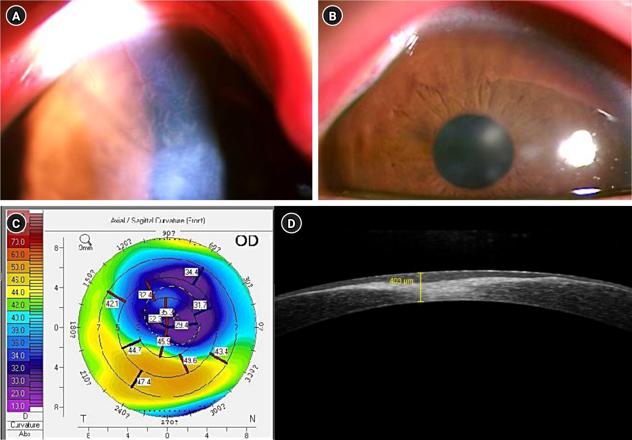
A 15-year-old male with XLA who had been receiving monthly IVIG was referred by a pediatrician for redness, epiphora, severe photophobia, and decreased vision in the right eye. He reported that his ocular symptoms recurred repeatedly, worsening approximately 1 week before each scheduled monthly IVIG infusion. At the initial visit, slit-lamp examination showed central corneal infiltration and corneal neovascularization (NV) extending from the superior cornea toward the center in the right eye, and multiple faint subepithelial opacities in the left eye (Fig. 1A, 1B). Uncorrected visual acuity (UCVA) was finger counting at 50 cm in the right eye and 0.2 in the left eye. In the right eye, corneal topography demonstrated superior corneal steepening, and anterior segment optical coherence tomography revealed a central corneal thinning of 403 µm and a hyperreflective lesion in the central corneal stroma (Fig. 1C, 1D).
Review of previous medical records showed that he had been diagnosed with XLA at 22 months old. At the time of diagnosis, the serum IgG level was <7.15 mg/dL (reference range, 700–1,600 mg/dL), IgA level was <6.2 mg/dL (reference range, 70–400 mg/dL), and IgM level was <17.5 mg/dL (reference range, 40–230 mg/dL). After IVIG treatment, IgG level reached the normal range, but IgA and IgM levels remained below the normal range. He first visited our clinic at 5 years of age due to redness in both eyes, at which time subepithelial corneal opacities in the left eye were observed. At 7 years of age, he revisited our clinic with redness and photophobia in both eyes, and bilateral corneal opacities were noted.
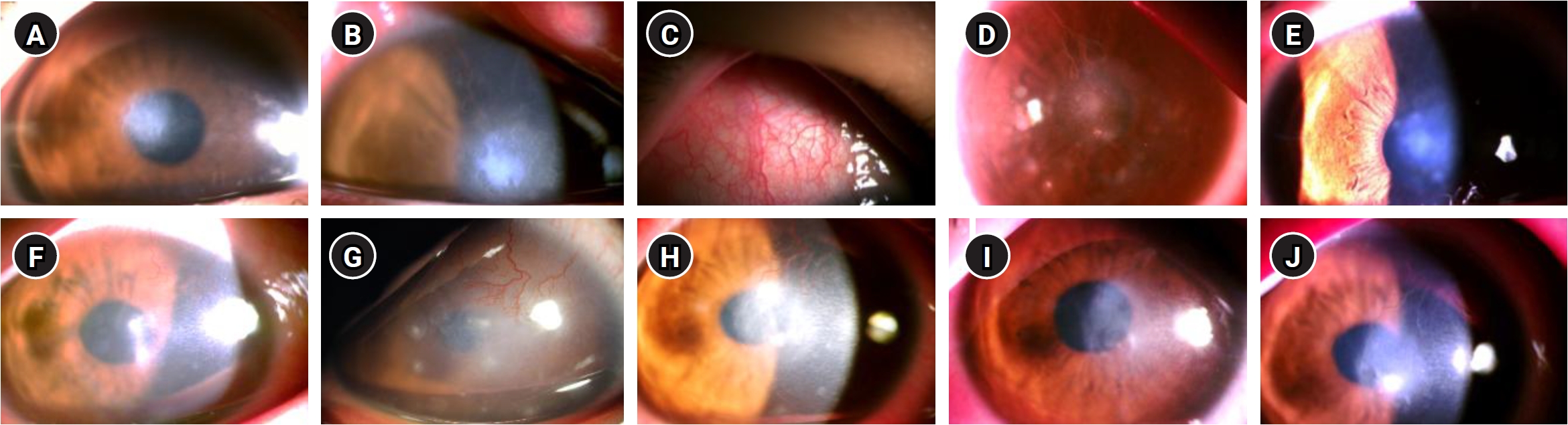
He was treated in the right eye with 0.3% ofloxacin and 0.1% fluorometholone eye drops four times daily, with weekly tapering. After 1 month, the corneal NV and central infiltration in the right eye had slightly decreased (Fig. 2A). However, 6 weeks after switching from 0.1% fluorometholone to 0.02% fluorometholone twice daily and then tapering to once daily, central infiltration increased (Fig. 2B). Steroid eye drops were changed to 0.1% fluorometholone again. Oral minocycline 50 mg was prescribed twice daily for 6 weeks, followed by once daily for an additional 3 months to treat blepharitis. Topical 0.1% fluorometholone was gradually tapered (twice daily for 2 weeks, once daily for 3 months, every other day for 2 months, and every 3 to 4 days for 4 weeks). To control recurrent episodes of transient conjunctival injection and itching in the right eye, 0.1% bromfenac and 0.05% epinastine hydrochloride eye drops were subsequently prescribed (Fig. 2C). After treatment, UCVA was 0.4 in the right eye and 0.5 in the left eye, and best-corrected visual acuity (BCVA) was 0.5 in the right eye and 1.0 in the left eye.
Six months later, he returned with redness and decreased vision in the left eye. BCVA in the left eye was 0.5 and did not improve with further refraction. Slit-lamp examination revealed superior corneal NV extending centrally, together with central corneal infiltration in the left eye (Fig. 2F). He was treated with 0.3% ofloxacin and 0.1% fluorometholone eye drops four times daily. After 1 week, the corneal infiltration had resolved, and both 0.3% ofloxacin and 0.1% fluorometholone were tapered to twice daily. He was scheduled for follow-up 2 weeks later but did not attend. Two months later, he returned without medication, complaining of severe photophobia, decreased vision, and conjunctival injection in the left eye. UCVA in the left eye was 0.04. Similar to the initial presentation in the right eye, severe central corneal infiltration and superior corneal NV were observed (Fig. 2G). Treatment with 1% prednisolone acetate four times daily and 0.3% ofloxacin every 3 hours for 1 week resulted in slight improvement of both the corneal infiltration and corneal NV (Fig. 2H). The 1% prednisolone acetate was subsequently tapered to three times daily for 2 weeks and changed to 0.5% loteprednol etabonate four times daily, which was gradually tapered over 4 weeks. At that time, UCVA was 0.5 in the right eye and 0.3 in the left eye. Thereafter, the patient again discontinued follow-up.
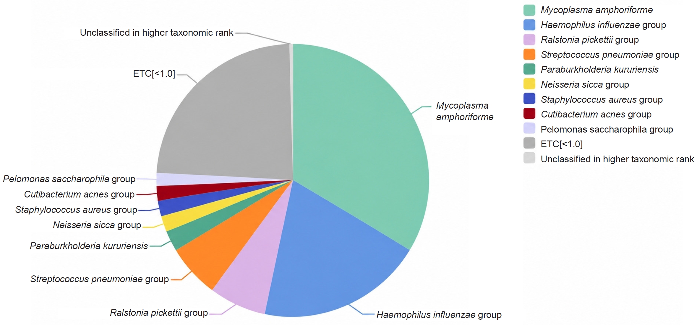
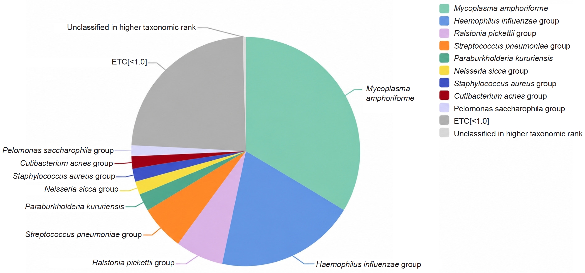
Eight months later, he returned for a routine examination showing central corneal opacity and superior ghost vessels in the right eye, and corneal opacity with thinning (minimum thickness: 309 µm) in the left eye (Fig. 2D, 2I). Swab specimens were obtained from the eyelids and conjunctivae of both eyes for bacterial culture and microbiome analysis approximately 6 months after all topical antibiotics had been discontinued. Bacterial culture identified H. influenzae and Staphylococcus epidermidis from the right eyelid, H. influenzae from the conjunctivae of both eyes, and no bacteria from the left eyelid. Microbiome analysis at the species level detected Mycoplasma amphoriforme (33.62%), the H. influenzae group (19.67%), the Ralstonia pickettii group (6.67%), the S. pneumoniae group (6.44%), Paraburkholderia kururiensis (2.44%), the Neisseria sicca group (1.93%), the S. aureus group (1.87%), the Cutibacterium acnes group (1.60%), and the Pelomonas saccharophila group (1.60%) in conjunctival specimens from both eyes (Fig. 3). Microbiome results were not obtained from the eyelid specimens. The patient was instructed to use 0.3% ofloxacin and 0.1% fluorometholone eye drops four times daily and to perform lid hygiene.
Swabs from both eyes were pooled into a single conjunctival specimen and analyzed by 16S rRNA gene sequencing (CJ Bioscience, Seoul, Korea). The V3–V4 region was sequenced on the Illumina MiSeq platform, and taxa were assigned against the EzBioCloud database (PKSSU4.0) at a 97% similarity cutoff for species-level identification. Of 6,430 valid reads, 6,284 (97.7%) were assigned to the species level; no negative (no-template) control was included.
At the 3-month follow-up visit, the central corneal opacity and corneal NV in both eyes had decreased (Fig. 2E, 2J). UCVA was 0.6 in the right eye and 0.15 in the left eye, and the patient was subsequently lost to follow-up again.
Ethics statement
This study was approved by the Institutional Review Board of Ewha Womans University Mokdong Hospital (IRB No. 2026-07-067) and was conducted in accordance with the tenets of the Declaration of Helsinki. The requirement for informed consent was waived owing to the retrospective nature of the study and the use of de-identified data.
Discussion
XLA is a primary immunodeficiency characterized by deficiency of B cells and immunoglobulins, predisposing affected patients to severe bacterial infections. The most common manifestations are upper respiratory tract infections, including otitis media, sinusitis, and pneumonia; gastrointestinal problems such as gastroenteritis and chronic diarrhea are also frequent [4]. Severe infections including sepsis, meningitis, and encephalitis have also been reported [4]. Among ophthalmic complications, recurrent keratoconjunctivitis has been described most frequently [4]. Similar to our case, a 32-year-old man with XLA was reported to have chronic progressive bilateral corneal disease with recurrent episodes, progressive corneal scarring, and visual loss during long-term follow-up [5]. These findings resemble those of phlyctenular keratoconjunctivitis, suggesting that immune dysregulation may play a role in chronic ocular surface inflammation in XLA [5,8]. Indeed, a recent immunologic study demonstrated that XLA involves not only B-cell deficiency but also altered T-cell subsets, supporting a broader mechanism for persistent ocular surface inflammation [8].
Previous studies have reported inconsistent therapeutic responses in keratoconjunctivitis associated with XLA. Mielke et al. [5] reported a 32-year-old man in whom IVIG was ineffective for conjunctivitis, whereas topical corticosteroid eye drops provided short-term symptomatic improvement and slowed the progression of corneal scarring. Abolhassani et al. [9] reported that IVIG reduced complications such as otitis media and pneumonia but not conjunctivitis or sinusitis. Kang et al. [6] reported a 15-year-old male with stromal keratitis and corneal thinning in whom the corneal lesion improved after IVIG treatment, but topical eye drops including corticosteroids did not improve the corneal lesions. Regarding corneal NV, Bock et al. [10] reported that topical bevacizumab eye drops (5 mg/mL, five times daily) reduced corneal NV in five patients. However, Mielke et al. [5] reported that subconjunctival bevacizumab at a concentration of 1.25 mg/0.05 mL produced no relevant effect after a single application.
In our case, topical corticosteroid treatment transiently improved corneal infiltration, opacity, and NV, but the disease recurred upon tapering or discontinuation of treatment. These recurrences characteristically occurred approximately 1 week before each scheduled IVIG infusion. This temporal pattern toward the end of the dosing interval is compatible with a decline in serum immunoglobulin levels before the next infusion. However, serum IgG was measured only at scheduled visits, not at the time of ocular exacerbations, and some values were obtained after periods of loss to follow-up. The available levels therefore do not reliably reflect the immunoglobulin status during symptomatic episodes. As this is a single case, no causal inference can be drawn. Residual IgA and IgM deficiency at mucosal surfaces, which IVIG replacement cannot restore, may also contribute to the persistent vulnerability of the ocular surface despite serum IgG replacement [3,4].
In our patient, conjunctival cultures identified H. influenzae, which is consistent with the report by Hansel et al. [7] who isolated H. influenzae from conjunctival swabs in three brothers with XLA. However, other studies evaluating causative organisms in XLA-associated keratoconjunctivitis have failed to isolate pathogens; Mielke et al. [5] reported that repeated conjunctival swabs were negative and Kang et al. [6] found negative corneal scraping cultures. To address these limitations of conventional culture, we performed microbiome analysis using culture-independent 16S rDNA sequencing to obtain a more comprehensive characterization of the ocular surface microbial environment [11].
Among the detected taxa, H. influenzae demonstrated the strongest correlation with both our culture results and the clinical presentation. Notably, M. amphoriforme accounted for the largest proportion of the microbiome (33.62%), a remarkable finding given its primary association with respiratory tract infections in immunocompromised patients. However, because it was detected solely by sequencing rather than culture, its direct pathogenic role remains to be determined.
To evaluate the clinical significance of the other identified organisms, we referenced prior studies on normal ocular surface flora. Ratnumnoi et al. [12] reported that coagulase-negative staphylococci and Streptococcus species may be part of the normal conjunctival and lid-margin flora. Jiang et al. [13] reported that the conjunctival sac flora of healthy children in East China was composed mainly of gram-positive cocci, most commonly S. epidermidis, S. aureus, and Streptococcus species. Based on these reference data, S. aureus, S. epidermidis, and C. acnes likely represent normal commensal flora. Among the remaining taxa, H. influenzae is the most plausible candidate pathogen, whereas the clinical significance of M. amphoriforme, S. pneumoniae, and N. sicca is uncertain. Ralstonia, Paraburkholderia, and Pelomonas species are commonly reported as reagent- or laboratory-derived contaminants in low-biomass 16S rRNA sequencing analyses and were therefore not regarded as clinically meaningful [12-14]. These findings suggest that, with further validation, microbiome profiling may help refine antibiotic selection in future management.
To our knowledge, this is the first report of conjunctival microbiome analysis in a patient with XLA. Microbiome analysis revealed a broader bacterial profile than culture alone, underscoring the value of culture-independent methods in immunodeficient patients. These findings suggest that microbiome profiling may, with further study, complement conventional culture in the management of persistent ocular surface inflammation in XLA.
This report has several limitations. First, this is a single case, and the microbiome analysis was based on a single sampling time point. Second, the microbiome result was derived from a single low-biomass conjunctival swab processed without a negative (no-template) control, so low-abundance taxa cannot be reliably distinguished from reagent- or laboratory-derived contamination [14]. Third, the specimens were obtained from the conjunctival surface rather than the cornea, so the findings indicate ocular-surface colonization rather than a proven causative role in the keratitis. Finally, 16S rRNA profiling identifies taxa but does not establish organism viability or antimicrobial susceptibility. In addition, serum IgG was not measured at the time of each ocular exacerbation.
Article Information
Author contributions
Conceptualization: BRK, KEH. Data curation: BRK. Investigation: BRK. Methodology: BRK, KEH. Project administration: KEH. Supervision: KEH. Formal analysis: BRK. Writing_original draft: BRK. Writing_review & editing: BRK, KEH.
Conflicts of interest
No potential conflict of interest relevant to this article was reported.
Funding
None.
Data availability
Not applicable.
Fig. 1.Anterior segment findings at the first visit. (A) Right-eye slit-lamp photograph showing central corneal infiltration and neovascularization extending centrally from the superior cornea. (B) Left-eye slit-lamp photograph showing multiple faint subepithelial opacities. (C) Right-eye corneal topography showing superior corneal steepening. (D) Right-eye anterior segment optical coherence tomography showing central corneal thinning to 403 µm and a hyperreflective central stromal lesion. OD, right eye.
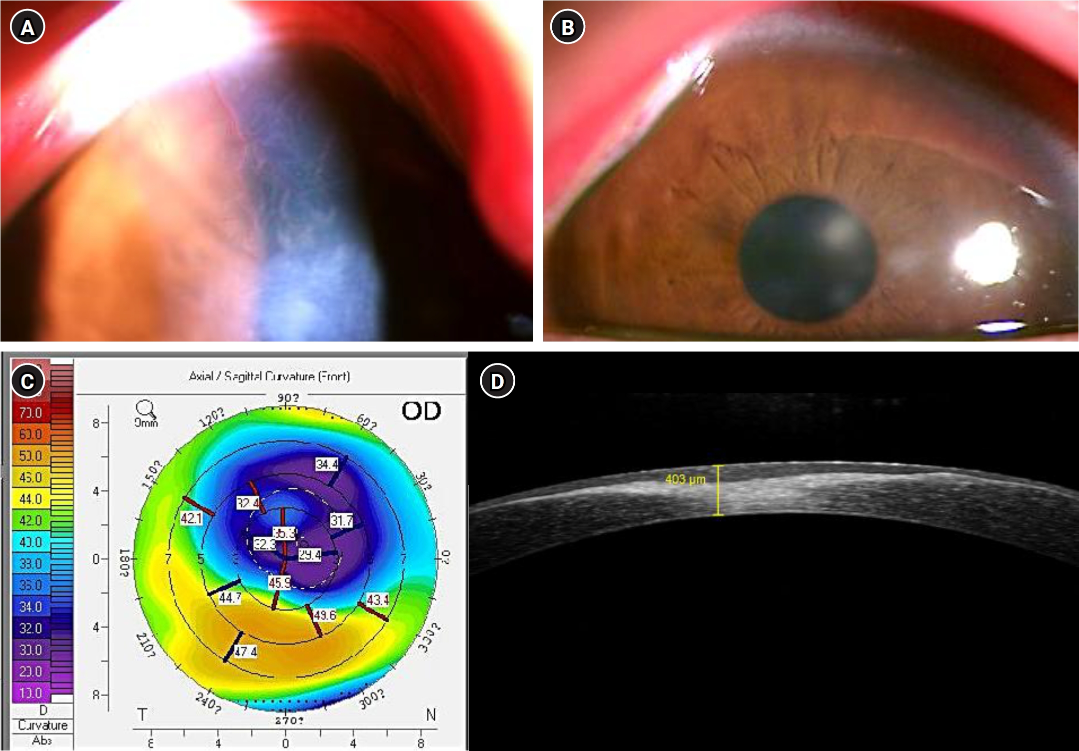
Fig. 2.Slit-lamp photographs of the right (A–E) and left (F–J) eyes during follow-up. (A) Regression of superior corneal infiltration and neovascularization after 1 month of treatment with 0.3% ofloxacin and 0.1% fluorometholone. (B) Recurrent superior corneal neovascularization and central infiltration after tapering corticosteroid eye drops. (C) Transient worsening of conjunctival injection during treatment. (D) Superior ghost vessels and multiple corneal opacities after loss to follow-up. (E) Residual corneal opacity and regression of superior neovascularization at the final follow-up. (F) New superior corneal neovascularization and central infiltration in the left eye 6 months later. (G) Worsening superior corneal neovascularization and severe central infiltration after 2 months without medication or follow-up. (H) Reduced corneal neovascularization and infiltration after treatment with 0.3% ofloxacin and 1% prednisolone acetate. (I) Central corneal opacities and thinning after loss to follow-up. (J) Residual corneal opacity and regression of neovascularization in the left eye at the final follow-up, similar to the right eye.
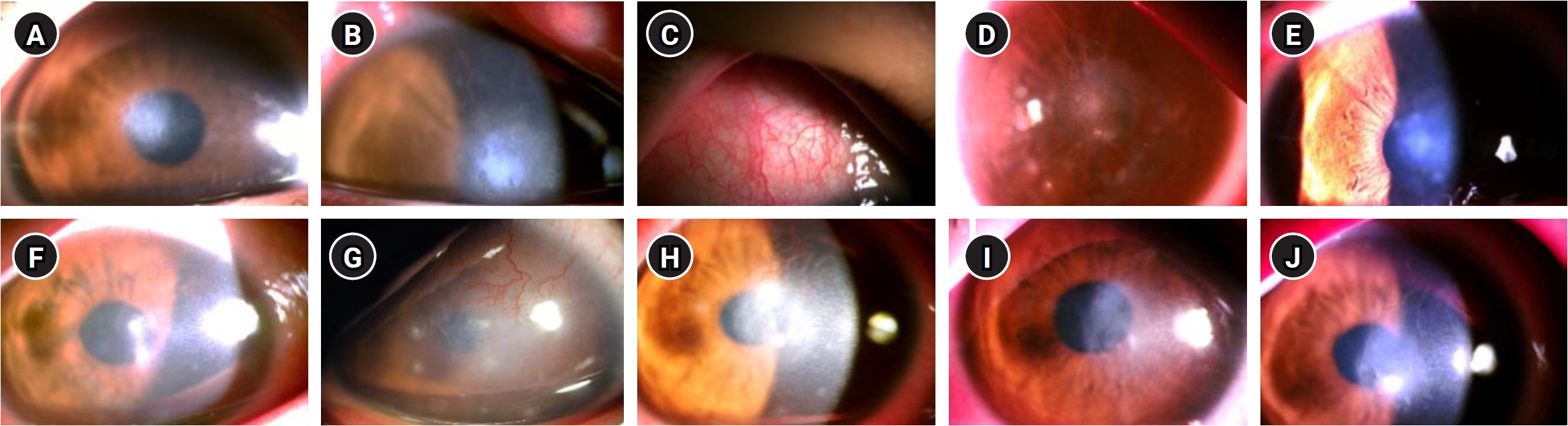
Fig. 3.Relative abundance of bacterial taxa in the conjunctival microbiome at the species level. Conjunctival swabs from both eyes were analyzed by sequencing the V3–V4 region of the 16S rRNA gene. Mycoplasma amphoriforme (33.62%) and the Haemophilus influenzae group (19.67%) were the most abundant taxa.
References
- 1. Cardenas-Morales M, Hernandez-Trujillo VP. Agammaglobulinemia: from X-linked to autosomal forms of disease. Clin Rev Allergy Immunol 2022;63:22‒35. ArticlePubMedPDF
- 2. Korea Disease Control and Prevention Agency (KDCA). 2023 Rare disease registry statistics [Internet]. Cheongju, Korea: KDCA; 2025 [cited 2026 Oct 1]. Available from: https://helpline.kdca.go.kr/cdchelp/ph/infNoti/selectAnnFrmDetail.do?menu=G0100&schNtbdCcd=01&ntbdSno=962.
- 3. Pieper K, Grimbacher B, Eibel H. B-cell biology and development. J Allergy Clin Immunol 2013;131:959‒71. ArticlePubMed
- 4. Winkelstein JA, Marino MC, Lederman HM, et al. X-linked agammaglobulinemia: report on a United States registry of 201 patients. Medicine (Baltimore) 2006;85:193‒202. ArticlePubMed
- 5. Mielke S, Grundel B, Schmidt SM, Tost F. Keratoconjunctivitis as a single entity in X-linked agammaglobulinemia? Ocul Immunol Inflamm 2023;31:665‒70. ArticlePubMed
- 6. Kang HJ, Cho YW, Yoo W, et al. Stromal keratitis in a patient with congenital hypogammaglobulinemia. J Korean Ophthalmol Soc 2020;61:554‒8. ArticlePDF
- 7. Hansel TT, O'Neill DP, Yee ML, et al. Infective conjunctivitis and corneal scarring in three brothers with sex linked hypogammaglobulinaemia (Bruton's disease). Br J Ophthalmol 1990;74:118‒20. ArticlePubMedPMC
- 8. Shelyakin PV, Lupyr KR, Egorov ES, et al. Naïve regulatory T cell subset is altered in X-linked agammaglobulinemia. Front Immunol 2021;12:697307.ArticlePubMedPMC
- 9. Abolhassani H, Hirbod-Mobarakeh A, Shahinpour S, et al. Mortality and morbidity in patients with X-linked agammaglobulinaemia. Allergol Immunopathol (Madr) 2015;43:62‒6. ArticlePubMed
- 10. Bock F, Konig Y, Kruse F, et al. Bevacizumab (Avastin) eye drops inhibit corneal neovascularization. Graefes Arch Clin Exp Ophthalmol 2008;246:281‒4. ArticlePubMedPDF
- 11. Zegans ME, Van Gelder RN. Considerations in understanding the ocular surface microbiome. Am J Ophthalmol 2014;158:420‒2. ArticlePubMedPMC
- 12. Ratnumnoi R, Keorochana N, Sontisombat C. Normal flora of conjunctiva and lid margin, as well as its antibiotic sensitivity, in patients undergoing cataract surgery at Phramongkutklao Hospital. Clin Ophthalmol 2017;11:237‒41. ArticlePubMedPMCPDF
- 13. Jiang M, Zhang J, Wan X, et al. Conjunctival sac flora and drug susceptibility analysis in normal children in East China. BMC Ophthalmol 2023;23:248.ArticlePubMedPMCPDF
- 14. Salter SJ, Cox MJ, Turek EM, et al. Reagent and laboratory contamination can critically impact sequence-based microbiome analyses. BMC Biol 2014;12:87.ArticlePubMedPMCPDF
Citations
Citations to this article as recorded by


 KSCRS



 ePub Link
 Cite
 XML Download