
Search
- Page Path
- HOME > Search
Original Article
- Comparison of combined torsional and conventional ultrasound mode versus torsional ultrasound mode phacoemulsification in different machines
- Jiyoung Emily Lee, Eun Chul Kim
- Insights Cataract Refract Surg 2026;11(2):40-46. Published online May 21, 2026
- DOI: https://doi.org/10.63375/icrs.25.018
-
 Abstract
 PDF
PDF ePub
ePub - Purpose
This study aimed to compare the clinical outcomes of phacoemulsification performed using a combined torsional and conventional ultrasound mode versus torsional ultrasound mode alone in cataract surgery.
Methods
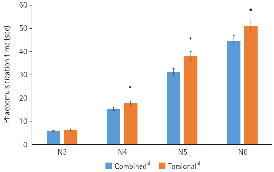
Sixty-five patients who underwent phacoemulsification using combined torsional and conventional ultrasound mode with the Cube alpha system (Nidek Co.) were compared with 300 patients who underwent phacoemulsification using torsional ultrasound mode alone with the Centurion system (Alcon Research Ltd.). The two groups were classified as N3, N4, N5, or N6 according to the Lens Opacities Classification System III. In each subgroup, phacoemulsification time (seconds), cumulative dissipated energy (CDE), balanced salt solution (BSS) use, and postoperative best-corrected visual acuity were analyzed and compared between the combined-mode group and the torsional-only group.
Results
In the N4–N6 subgroups, the torsional-only group had significantly longer phacoemulsification times and higher CDE values than the combined-mode group (P<0.05 for all comparisons). In the N3–N6 subgroups, the total volume of BSS used in the torsional-only group (N3, 30.97±11.05; N4, 48.56±20.06; N5, 68.27±23.06; N6, 125.64±45.67) was significantly greater than that used in the combined-mode group (N3, 23.01±10.27; N4, 39.03±24.16; N5, 42.86±18.24; N6, 54.75±0.75) (P<0.05). There were no statistically significant differences between the groups in postoperative manifest refraction spherical equivalent, corneal endothelial cell loss, or best-corrected visual acuity.
Conclusion
Compared with torsional ultrasound mode alone, the combined torsional and conventional ultrasound mode was more efficient in terms of ultrasound time, CDE, and BSS use. This advantage was more evident with increasing nuclear sclerosis grade.
- 432 View
- 4 Download

Review Articles
- Phacoemulsification in patients with diabetes: from preoperative evaluation to postoperative management
- Yeoun Sook Chun
- Insights Cataract Refract Surg 2025;10(3):65-75. Published online October 31, 2025
- DOI: https://doi.org/10.63375/icrs.25.012
-
Abstract
PDFePub
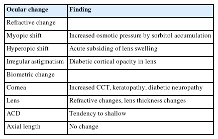
- Diabetes mellitus is one of the most common chronic diseases worldwide and is a leading cause of blindness in patients over the age of 50 years. Patients with diabetes have an elevated risk of developing cataracts compared to individuals without diabetes; furthermore, cataracts also tend to progress more rapidly in this population, leading to the need for surgery at a younger age. This review aims to summarize the key considerations in the management of cataract surgery in patients with diabetes, from preoperative evaluation to postoperative care. Patients with diabetes often present with unstable refractive status, dry eye disease, corneal epithelial defects, and recurrent corneal erosions. They also tend to have reduced corneal endothelial cell density and small pupils, both of which increase the risk of intraoperative complications. Postoperatively, these patients are at risk of developing pseudophakic cystoid macular edema, posterior capsular opacification, endophthalmitis, progression of diabetic retinopathy, and neovascular glaucoma. Patients with long-standing or poorly controlled diabetes face a higher likelihood of postoperative complications, highlighting the importance of regular ophthalmic follow-up examinations. Furthermore, adjunctive treatments such as timely intravitreal injections of anti-vascular endothelial growth factor agents may reduce the risk of vision-threatening complications following cataract surgery.
- 1,261 View
- 14 Download
- Trabecular microbypass using iStent combined with cataract surgery
- Yeoun Sook Chun
- Insights Cataract Refract Surg 2025;10(2):42-51. Published online June 30, 2025
- DOI: https://doi.org/10.63375/icrs.25.007
-
Abstract
PDFePub
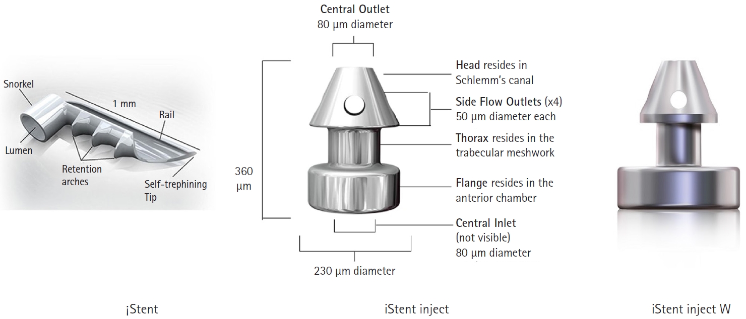
- Minimally invasive glaucoma surgery has revolutionized conventional glaucoma treatment due to its simple procedures, rapid recovery, and few complications. iStent, a trabecular microbypass that can be implanted via combined cataract surgery, has the advantage of lowering intraocular pressure (IOP) independent of bleb formation. Furthermore, it is straightforward to implement and does not involve a substantial burden. iStent has a synergetic effect with phacoemulsification. Combining iStent with phacoemulsification yielded a greater reduction of IOP and glaucoma eyedrops, as well as higher rates of visual field change, than when either modality was used in isolation. iStent has emerged as a new treatment option for patients with mild to moderate glaucoma. This review aims to improve readers’ understanding of iStent by summarizing the surgical techniques needed to correctly insert it for optimal outcomes and discussing problem-solving in the context of patient care.
- 1,510 View
- 10 Download
Original Article
- Clinical manifestations after cataract surgery in patients with moderate Fuchs corneal endothelial dystrophy
- Myung-Sun Song, Dong Hyun Kim
- Insights Cataract Refract Surg 2025;10(1):19-25. Published online February 28, 2025
- DOI: https://doi.org/10.63375/icrs.25.006
-
Abstract
PDFePub
- Purpose
The aim of this study was to analyze the clinical outcomes of cataract surgery in patients with moderate Fuchs endothelial corneal dystrophy (FECD) in whom central endothelial cells could not be observed using specular microscopy.
Methods
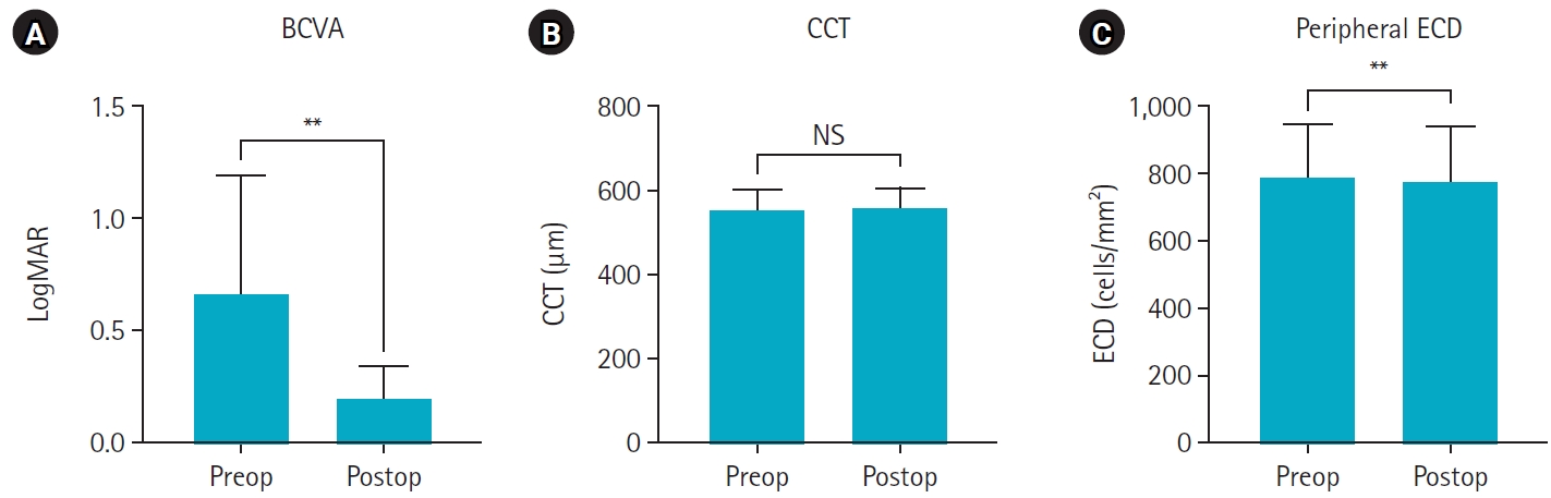
This retrospective study included nine eyes in seven patients diagnosed with FECD who underwent phacoemulsification at a single institution between January 2023 and November 2024. A single experienced corneal specialist performed slit-lamp examination and phacoemulsification. Best-corrected visual acuity (BCVA), specular microscopy, and central corneal thickness (CCT) measurements were performed preoperatively and postoperatively, and the outcomes were compared.
Results
The mean age of the patients was 69.8±6.5 years. Three were male patients and four were female patients. The mean preoperative CCT was 559.5±51.8 μm and the mean peripheral endothelial cell density was 599.3±129.4 cells/mm2. BCVA significantly improved in all patients postoperatively, with a mean logMAR BCVA improving from 0.65±0.52 preoperatively to 0.19±0.14 postoperatively (P=0.002). The mean CCT showed no significant change (preoperative, 559.6±51.8 μm; postoperative, 566.8±45.1 μm; P=0.218). In patients with follow-up longer than 6 months, an increase in CCT was observed at 30 days postoperatively, but CCT returned to preoperative levels after 90 days.
Conclusion
In patients with moderate-to-severe FECD in whom central endothelial cells cannot be measured, phacoemulsification may provide favorable visual outcomes if peripheral endothelial cells are observed and corneal edema is absent preoperatively.
- 1,344 View
- 14 Download
 First
First Prev
Prev

