-
Utilization of intraoperative optical coherence tomography during Descemet stripping automated endothelial keratoplasty: a case report
-
Ji Hyun Yoon, Ho Sik Hwang
-
Received April 29, 2026 Accepted June 13, 2026 Published online July 3, 2026
-
DOI: https://doi.org/10.63375/icrs.26.004
-
-
 Abstract Abstract
 ePub ePub
- Purpose
We report a case demonstrating the clinical utility of real-time intraoperative anterior-segment optical coherence tomography (iOCT) for visualizing interface-fluid drainage through venting incisions during Descemet stripping automated endothelial keratoplasty (DSAEK) in a patient with bullous keratopathy.
Case
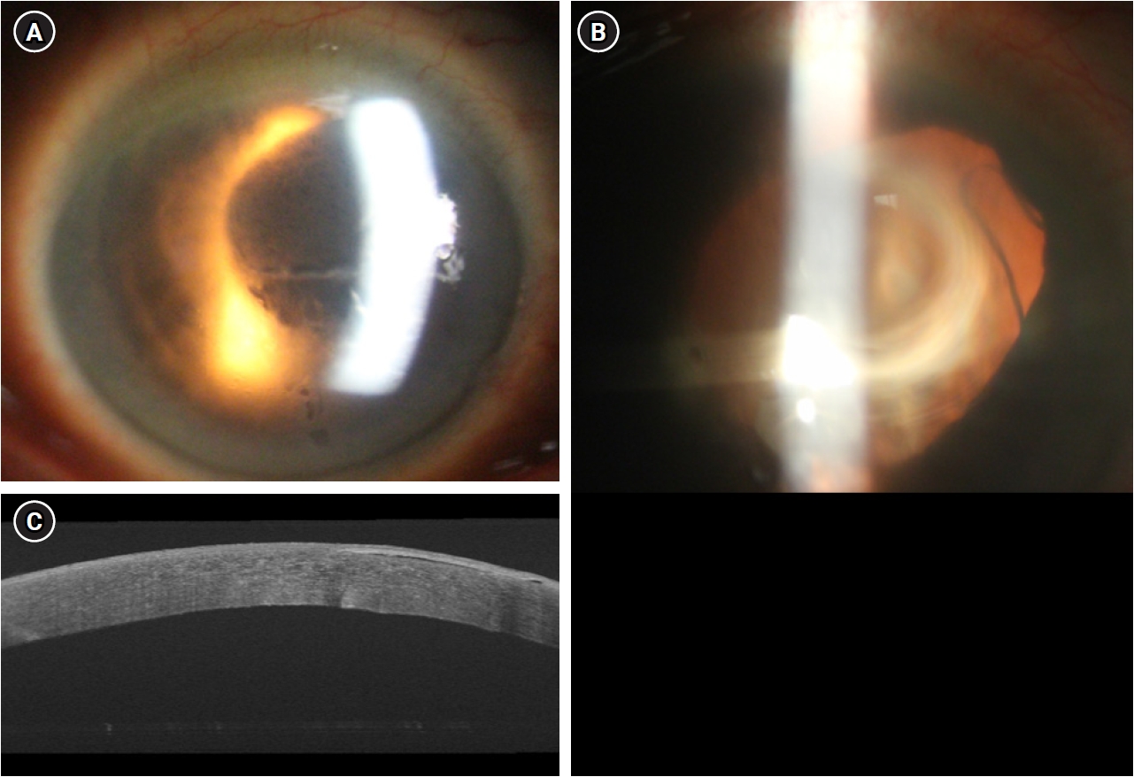
summary: A 77-year-old male patient with a history of bilateral cataract surgery presented with a several-month history of decreased vision in the right eye. Slit-lamp examination revealed corneal edema with bullae, and central corneal thickness was 700 µm. The endothelial cell count could not be measured by specular microscopy. With a diagnosis of pseudophakic bullous keratopathy, DSAEK was performed. After graft insertion and anterior-chamber air tamponade, iOCT identified residual interface fluid between the host stroma and donor graft. Venting incisions were then created, and the residual fluid was expressed with a sponge. Real-time iOCT confirmed the gradual disappearance of the interface fluid and complete apposition of the graft to the host cornea. After surgery, the graft remained stably attached without rejection or detachment during 3 months of follow-up.
Conclusion
During DSAEK, iOCT allows immediate verification of interface-fluid removal through venting incisions and may help improve graft positioning and early adherence.
|

 KSCRS
KSCRS